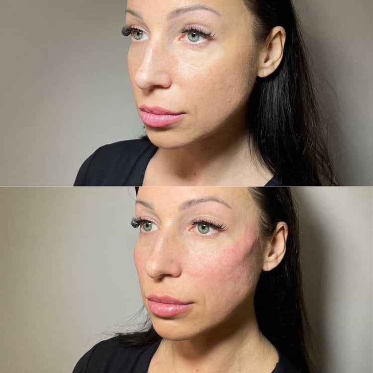
Profhilo’s remarkable success in the aesthetics industry can be attributed to its unique composition and multifaceted approach to skin rejuvenation.
At its core, Profhilo is a biorevitalization treatment comprised of hyaluronic acid (HA) – a naturally occurring substance found abundantly in our bodies. HA acts as a powerful humectant, attracting and retaining moisture, thereby plumping the skin and restoring its youthful hydration.
However, Profhilo differentiates itself by incorporating a unique concentration and cross-linking structure of HA. This specialized formulation allows for a sustained release of HA over time, leading to prolonged hydration and collagen stimulation.
Beyond its high-quality HA, Profhilo stands out for its innovative delivery method. Unlike traditional dermal fillers that are injected superficially, Profhilo is administered via a specific technique known as the “Profhilo injection protocol.” This involves strategically placing multiple tiny injections throughout the treatment area, creating a web-like distribution of HA beneath the skin.
This unique placement maximizes tissue integration and stimulates fibroblasts, the cells responsible for producing collagen and elastin – the essential proteins that provide skin structure and elasticity.
The result is a multi-pronged approach to rejuvenation: increased hydration, boosted collagen production, improved skin tone and texture, and enhanced overall radiance. This holistic effect sets Profhilo apart from other dermal fillers and treatments, making it a popular choice for those seeking natural-looking, long-lasting improvements.
Profhilo’s success is a testament to its innovative composition and effective treatment protocol, offering a comprehensive solution to address various signs of skin aging.
Clinical Evidence for Efficacy
Profhilo’s success stems from a unique combination of factors that address multiple aspects of skin health.
Profhilo utilizes a high-concentration, stabilized hyaluronic acid (HA) gel.
*
This HA is specifically cross-linked with BDDE, resulting in longer-lasting hydration and volume restoration compared to traditional HA fillers.
2. Bioremodel® Technology:
*
Profhilo’s unique “Bioremodel®” technology involves a precise network of microscopic needles that distribute the HA gel within the skin layers.
*
This method triggers a cascade of biological responses, including collagen and elastin production, leading to improved skin texture, firmness, and elasticity.
3. Versatility in Applications:
*
Profhilo can be effectively used for various concerns, including:
Contact Us
It’s Me and You Clinic – Anti-Wrinkle, Dermal Filler and Skincare Clinic, Kingston, Surrey
Neck and décolletage tightening – addressing laxity and sagging skin
*
Hand rejuvenation – restoring volume and smoothing out veins
4. Minimal Downtime:
*
Profhilo treatments are generally well-tolerated with minimal downtime.
*
Patients typically experience mild redness or swelling that resolves within a few days.
5. Clinically Proven Results:
*
Numerous clinical studies have demonstrated Profhilo’s efficacy in improving skin hydration, elasticity, and reducing the appearance of wrinkles.
*
These studies show significant improvement in skin quality compared to placebo or other treatments.
While alternatives exist for facial rejuvenation and skin tightening, Profhilo’s unique combination of HA formulation, Bioremodel® technology, versatility, minimal downtime, and strong clinical evidence contributes to its widespread success and popularity among both practitioners and patients.
Exploring Alternative Treatments
Dermal Fillers: A Different Approach
While Profhilo has gained significant popularity for its skin-rejuvenating effects, it’s essential to remember that individual needs and preferences vary greatly. Exploring alternative treatments can be beneficial to determine what best suits your specific concerns and desired outcomes.
Dermal fillers, a popular category of injectables, offer a diverse range of options beyond Profhilo. They work by adding volume to targeted areas, plumping up wrinkles, and restoring lost facial contours. Unlike Profhilo, which primarily focuses on stimulating collagen production, fillers provide immediate results.
Hyaluronic acid-based fillers are among the most commonly used. These gels attract and retain moisture, providing a natural-looking hydration boost and smoothing out fine lines and wrinkles. Some popular hyaluronic acid fillers include Juvederm, Restylane, and Belotero.
Calcium hydroxylapatite (CaHA) fillers offer another alternative. They are designed to stimulate collagen growth, resulting in gradual improvements in skin texture and elasticity. Radiesse is a well-known CaHA filler.
For individuals seeking longer-lasting results, poly-L-lactic acid (PLLA) fillers can be considered. They gradually degrade over time, prompting the body to produce new collagen and providing a sustained lifting effect. Sculptra is an example of a PLLA filler.
When choosing a dermal filler, it’s crucial to consult with a qualified and experienced injector. They will assess your skin type, concerns, and desired outcome to recommend the most suitable option and ensure safe and effective treatment.
Besides Profhilo and dermal fillers, other non-surgical skin rejuvenation techniques include microneedling, laser treatments, chemical peels, and thread lifts. Each approach has its own benefits and considerations, making a thorough evaluation essential for finding the best fit.
Microneedling with PRP: Stimulating Natural Collagen Production
Microneedling with platelet-rich plasma (PRP) has emerged as a popular alternative treatment for skin rejuvenation, offering potential benefits comparable to injectables like Profhilo.
Both Profhilo and microneedling with PRP stimulate collagen production, leading to improved skin elasticity, firmness, and texture. However, their mechanisms of action differ.
Profhilo is a hyaluronic acid-based injectable that works by hydrating the skin and attracting water molecules. This plumps up the dermis, smoothing wrinkles and improving overall skin tone.
Microneedling with PRP, on the other hand, involves using tiny needles to create controlled micro-injuries in the skin. These injuries trigger a natural healing response, stimulating the production of collagen, elastin, and other growth factors.
PRP is derived from the patient’s own blood, which is processed to concentrate platelets—cells rich in growth factors that promote tissue repair and regeneration.
When PRP is combined with microneedling, the growth factors are directly delivered into the deeper layers of the skin, enhancing collagen production and accelerating healing.
One advantage of microneedling with PRP is its versatility. It can be customized to address various skin concerns, including wrinkles, acne scars, uneven skin tone, and stretch marks.
Another benefit is that it is less invasive than surgical procedures and carries a lower risk of complications compared to injectables.
However, microneedling with PRP may require multiple sessions to achieve optimal results, and downtime can be longer than with Profhilo.
The choice between Profhilo and microneedling with PRP depends on individual skin type, concerns, and preferences. Some patients may prefer the quick and easy results of Profhilo, while others may opt for the long-term benefits and natural collagen stimulation offered by microneedling with PRP.
The Verdict: Finding the Right Solution for You
Individualized Needs and Goals
While Profhilo has gained significant popularity for its hyaluronic acid-based, skin rejuvenation properties, it’s important to remember that *individual needs and goals* vary greatly.
What works wonders for one person might not yield the same results for another. Therefore, seeking a solution tailored to your specific requirements is crucial.
Consider these factors when exploring alternatives to Profhilo:
**Skin Concerns:** What specific skin concerns are you addressing? Are you looking to reduce wrinkles, improve skin hydration, enhance elasticity, or address uneven texture?
**Desired Outcome:** What do you hope to achieve with your treatment? Do you want subtle rejuvenation or a more dramatic transformation?
**Skin Type:** Different skin types react differently to various treatments. A professional can assess your skin type and recommend suitable options.
**Budget:** Treatment costs vary significantly. Determine your budget range to narrow down your choices.
**Downtime:** Some treatments require more downtime than others. Consider your lifestyle and how much time you’re willing to dedicate to recovery.
Here are some popular alternatives to Profhilo:
* **Fillers:** Hyaluronic acid fillers like Restylane and Juvederm can address specific concerns such as wrinkles, volume loss, and lip enhancement.
* **Botox:** Botulinum toxin injections can temporarily paralyze muscles, smoothing wrinkles and preventing new ones from forming.
* **Microneedling:** This minimally invasive procedure uses fine needles to stimulate collagen production, improving skin texture and reducing the appearance of scars.
* **Chemical Peels:** Chemical peels exfoliate the skin, revealing a smoother, brighter complexion. Different peel strengths cater to various skin concerns.
* **Laser Treatments:** Various laser technologies target specific skin concerns, such as pigmentation, redness, and wrinkles.
Consulting with a qualified and experienced dermatologist or aesthetic practitioner is essential for determining the most suitable treatment for your individual needs and goals. They can provide personalized recommendations based on your skin type, desired outcome, and lifestyle.
Consultation with a Qualified Professional
Profhilo has gained significant popularity as a hyaluronic acid-based dermal filler, renowned for its hydrating and skin-rejuvenating properties.
However, the quest for optimal skincare solutions often leads individuals to explore alternatives.
While Profhilo offers compelling benefits, it might not be the perfect fit for everyone or every concern.
Consulting a qualified professional is crucial to determine the most suitable treatment for your individual needs and goals.
Understanding Your Skin Concerns:
The first step involves identifying your specific skin concerns. Are you seeking hydration, wrinkle reduction, volume restoration, or a combination of these? A qualified professional can assess your skin type, condition, and desired outcomes.
Beyond Profhilo, various other treatments exist that target different aspects of skin aging. These include:
Hyaluronic Acid Fillers:
Similar to Profhilo, but with varying formulations and injection techniques, offering customizable results for specific concerns.
Collagen Stimulators:**
Substances like Sculptra and Radiesse stimulate collagen production, providing long-lasting volume and skin tightening effects.
Microneedling:
Uses tiny needles to create controlled micro-injuries, prompting the skin’s natural healing process and collagen synthesis.
Laser Treatments:
Different lasers target specific concerns like pigmentation, texture irregularities, and fine lines.
Individualized Assessment:
A qualified professional will carefully analyze your skin, medical history, and desired results to recommend the most appropriate treatment plan.
Realistic Expectations:**
It’s important to have realistic expectations about the outcomes of any aesthetic treatment. A qualified professional will discuss potential benefits, limitations, and possible side effects.
Safety and Expertise:
Always choose a licensed and experienced medical professional who uses safe and effective techniques and products.
Ultimately, the best solution for you may not necessarily be Profhilo. A thorough consultation with a qualified professional will empower you to make an informed decision that aligns with your individual needs and aesthetic aspirations.
Tear trough filler is a popular cosmetic procedure used to address under-eye hollows, creating a more youthful and refreshed appearance. It involves injecting dermal fillers into the tear trough area – the hollow beneath the eye.
However, sometimes patients may experience dissatisfaction with the results, desire a change in their appearance, or develop complications like lumps or unevenness. In these cases, dissolving tear trough filler becomes necessary.
The process involves using an enzyme called hyaluronidase. Hyaluronidase breaks down hyaluronic acid, the primary ingredient in many dermal fillers. This effectively reverses the effects of the filler, allowing it to be reabsorbed by the body.
Understanding the different types of fillers used in tear trough treatment is crucial before considering dissolution.
Contact Us
It’s Me and You Clinic – Anti-Wrinkle, Dermal Filler and Skincare Clinic, Kingston, Surrey
Kingston upon Thames, Surrey, United KingdomKT2 6LX
Hyaluronic Acid Fillers:
These are the most common type of filler used in tear troughs due to their natural properties and reversible nature. Popular examples include:
Juvederm
Restylane
Belotero
These fillers are made from hyaluronic acid, a naturally occurring substance in the skin that attracts and holds moisture.
They provide volume to the under-eye area, smoothing out hollows and reducing the appearance of dark circles.
Dissolving these fillers with hyaluronidase is straightforward due to their compatibility with this enzyme.
Calcium Hydroxyapatite (CaHA) Fillers:
These fillers offer a more permanent solution compared to hyaluronic acid fillers. They are composed of microscopic calcium crystals suspended in a gel. CaHA fillers can provide longer-lasting results, but dissolution with hyaluronidase is not as effective.
If a patient desires to dissolve CaHA filler, a different approach may be necessary, involving techniques like manual removal or laser treatments.
Reasons for Dissolution
Dissolving tear trough filler involves using an enzyme called hyaluronidase to break down hyaluronic acid fillers, which are commonly used to treat under-eye hollows.
Hyaluronic acid is a naturally occurring substance in the body that attracts and holds water, giving the skin its plumpness and hydration.
Tear trough fillers utilize this property to restore volume and smooth out wrinkles under the eyes.
However, as with any cosmetic procedure, there are instances where dissolving tear trough filler becomes necessary.
Here are some reasons why someone might choose to dissolve tear trough filler:
Dissatisfaction with Results: The most common reason for dissolving filler is simply not being happy with the outcome. This could include the filler looking unnatural, too puffy, or placed incorrectly.
Allergic Reaction:**
A small percentage of people may experience an allergic reaction to hyaluronic acid fillers.
Symptoms can range from mild redness and swelling to more severe reactions that require medical attention.
Migration or Lumping:**
In some cases, the filler can migrate away from the intended area or form lumps that are visible or uncomfortable.
Dissolving the filler can correct this issue.
Changes in Desired Look: People’s aesthetic preferences change over time. They may decide they no longer want the fullness provided by the tear trough filler, or their facial structure may naturally evolve, requiring adjustments to their filler placement.
It is important to remember that dissolving tear trough filler is a medical procedure that should only be performed by a qualified and experienced injector.
They will assess your individual situation, discuss the risks and benefits, and determine the best course of action for you.
The Dissolution Process
Choosing a Dissolving Agent
Dissolution of tear trough fillers involves the enzymatic breakdown of hyaluronic acid (HA) filler material using a specific dissolving agent. Hyaluronic acid is a natural substance found in our skin that helps maintain hydration and volume. However, when used as a dermal filler, its effects are temporary, typically lasting 6 months to a year.
The most common method for dissolving tear trough fillers is with hyaluronidase. This enzyme breaks down the HA molecules in the filler, allowing them to be absorbed by the body. Hyaluronidase is injected directly into the area of the filler, and it begins to work almost immediately. Most patients experience a noticeable softening of the filler within minutes to hours.
When choosing a dissolving agent for tear trough fillers, several factors need to be considered:
**Type of Filler:** Different HA fillers may have varying compositions and molecular weights, which can affect their susceptibility to hyaluronidase. The practitioner should know the specific type of filler used during the initial injection.
**Patient’s Medical History:** Allergies or sensitivities to medication should be carefully evaluated. It’s important to avoid using a dissolving agent that could trigger an adverse reaction.
**Location and Amount of Filler:** The amount and location of the filler will influence the dosage and technique used for dissolution.
Hyaluronidase is generally considered safe and effective for dissolving tear trough fillers. However, there are potential risks and side effects associated with any injection procedure:
* Bruising or swelling: These are common side effects that typically resolve within a few days.
* **Redness or irritation:** This can occur at the injection site and usually subsides on its own.
* **Allergic reaction:** Although rare, some people may experience an allergic reaction to hyaluronidase.
It’s essential to choose a qualified and experienced injector who understands the nuances of filler dissolution. They will be able to assess your individual needs and determine the most appropriate dissolving agent and technique for your situation.
Administration Technique
Dissolving tear trough filler involves strategically breaking down hyaluronic acid (HA) fillers that have been injected under the eyes to address undereye hollows or dark circles. This procedure is typically performed by a qualified medical professional, such as a dermatologist or plastic surgeon, who has experience with injectables and facial anatomy.
The primary tool used in the dissolution process is hyaluronidase, an enzyme that specifically breaks down hyaluronic acid molecules. Hyaluronidase comes in various formulations, including both injectable and topical preparations.
During the procedure, a small amount of hyaluronidase is carefully injected into the area where the filler was previously placed. The enzyme works by hydrolyzing (breaking apart) the HA chains, gradually dissolving the filler material over time.
The administration technique typically involves:
1. **Cleaning and Anesthesia:** The treatment area is thoroughly cleansed, and topical anesthesia may be applied to minimize any discomfort.
2. **Injection Technique:** Using a fine needle, the hyaluronidase is injected into multiple small points within the filler area. This ensures even distribution of the enzyme and promotes efficient breakdown of the HA filler.
3. **Massaging:** After injection, gentle massage may be performed to help distribute the hyaluronidase further and facilitate the dissolution process.
4. **Observation:** The medical professional will monitor the area closely for any immediate reactions or side effects.
The time required for complete filler dissolution varies depending on factors such as the amount of filler used, individual metabolism, and the specific hyaluronidase formulation. It may take several days to a couple of weeks for the filler to fully dissolve.
Following the procedure, patients may experience some mild swelling, bruising, or redness. These side effects typically subside within a few days.
It is essential to consult with a qualified medical professional to discuss your individual needs and expectations regarding tear trough filler dissolution. They can assess your suitability for the procedure, determine the appropriate technique, and provide guidance on post-treatment care.
Post-Dissolution Care
Managing Swelling and Bruising
Post-dissolution care focuses on minimizing any residual effects after dissolving tear trough filler, promoting healing, and restoring a natural appearance.
Immediately following the procedure, ice packs can be applied to reduce swelling and discomfort. Gently massaging the treated area can also help improve circulation and minimize bruising.
In the first few days after dissolution, it’s crucial to avoid touching or rubbing the eyes excessively. Sleeping with your head elevated can further help manage swelling.
Use gentle, fragrance-free skincare products formulated for sensitive skin to cleanse the area and avoid irritation. Sun exposure should be minimized as the skin may be more sensitive during healing.
Hydration is key to supporting the body’s natural healing process. Drinking plenty of water can help flush out toxins and promote cell regeneration.
Swelling typically subsides within a few days to a week, while bruising may last longer, sometimes up to two weeks. If you experience excessive swelling, prolonged bruising, or any signs of infection, consult your practitioner immediately.
Avoid strenuous activities, makeup, and contact lenses for the first few days after treatment. Makeup can irritate the delicate skin and delay healing.
Expected Results and Timeline
Post-dissolution care involves maintaining proper skin hygiene and minimizing irritation to promote healing and prevent complications.
Patients are typically advised to avoid direct sunlight, intense heat, or cold exposure for a few days after the procedure. Sun protection with a broad-spectrum sunscreen is crucial during the recovery period.
Gentle cleansing with mild soap and water can be used twice daily to maintain skin hygiene. Harsh scrubs or exfoliants should be avoided until the treated area has fully healed.
Makeup application is usually safe after 24 hours, but it’s important to use non-irritating products and avoid rubbing the treated area.
Expected results of tear trough filler dissolution include a gradual reduction in the appearance of dark circles or hollows under the eyes. The extent of improvement depends on factors like the amount of filler injected, individual skin characteristics, and post-treatment care.
The timeline for complete results can vary widely from person to person, typically ranging from a few weeks to several months. During this period, some temporary swelling or redness may be noticeable as the body absorbs the dissolved filler.
It’s essential to consult with a qualified medical professional to discuss individual expectations, risks, and post-procedure care instructions. Regular follow-up appointments can help monitor healing progress and address any concerns that may arise.
Several factors influence how long you need to wait between lip filler appointments, as everyone’s body heals at a different pace.
Here are some key considerations:
Amount and Type of Filler Used: Larger volumes or thicker fillers may require a longer downtime for optimal results.
Technique Employed by the Injector: The injection technique used can affect bruising, swelling, and overall healing time.
Individual Healing Rate: Genetics, age, skin type, and overall health all play a role in how quickly your body recovers.
Some individuals may experience minimal bruising or swelling and feel comfortable getting lip filler again within two weeks. Others might need four to six weeks for their lips to fully heal before a second treatment.
It’s important to consult with your injector to determine the appropriate waiting period for you. They can assess your individual healing characteristics, consider the filler type used, and provide personalized guidance on when it’s safe to have additional lip filler.
Generally, aiming for a wait time of at least four weeks between treatments is a good rule of thumb. However, always prioritize your body’s needs and listen to your injector’s recommendations.
Type of Filler Used
Numerous factors influence the recommended interval between lip filler injections. These factors can be broadly categorized as patient-specific and product-specific.
Patient-specific factors encompass individual metabolism, skin elasticity, lifestyle choices, and desired outcome. Individuals with faster metabolisms may see their filler dissolve quicker, requiring more frequent touch-ups. Conversely, those with slower metabolisms might experience longer-lasting results.
Skin elasticity plays a crucial role; more elastic skin tends to hold onto filler better, extending the time between treatments. Lifestyle factors like sun exposure and smoking can accelerate filler breakdown, necessitating shorter intervals.
The desired outcome also impacts timing. For subtle enhancement, longer intervals might be appropriate, while achieving dramatic volume requires more frequent touch-ups.
Product-specific factors include the type of hyaluronic acid filler used. Different formulations have varying molecular weights and viscosities, affecting their longevity and how they integrate with the natural tissue.
Hyaluronic acid (HA) fillers are broadly classified into those designed for fine lines and wrinkles and those formulated for volume augmentation.
Fine line HA fillers tend to be more rapidly absorbed, requiring touch-ups every 3-6 months. Volume-enhancing HA fillers, with their larger particle sizes, typically last 6-18 months or longer.
Moreover, newer hyaluronic acid technologies boast cross-linking, which creates stronger bonds between molecules, enhancing longevity and potentially extending the time between injections to up to 2 years.
Desired Results
Several factors influence the recommended interval between lip filler appointments. The primary factor is the type of filler used.
Hyaluronic acid fillers, which are the most common type, tend to last between 6-18 months depending on individual metabolism and lifestyle.
Dermal fillers made from other substances like poly-L-lactic acid can last longer, up to two years or more.
Another crucial factor is the desired results. A patient seeking subtle enhancement might opt for less frequent touch-ups, potentially every 12 months.
Those desiring a fuller, more dramatic look may need to schedule appointments every 6-8 weeks initially, with subsequent sessions spaced further apart as their lips settle.
Lip movement and expression also play a role. Individuals who frequently move their lips while talking or smiling might experience quicker filler breakdown and require more frequent touch-ups.
Lifestyle factors such as sun exposure, smoking, and hydration can impact the longevity of lip fillers.
Contact Us
It’s Me and You Clinic – Anti-Wrinkle, Dermal Filler and Skincare Clinic, Kingston, Surrey
Excessive sun exposure can break down hyaluronic acid molecules faster, requiring more frequent maintenance.
Smoking dehydrates the skin and compromises blood flow, hindering filler absorption and longevity.
Staying adequately hydrated promotes collagen production, helping to support and maintain the fullness achieved with lip fillers.
General Guidelines
Initial Touch-Ups
Determining the optimal timeframe between lip filler appointments depends on several factors, including individual healing rates, desired volume, and the type of filler used.
Here are some general guidelines:
Initial Touch-Ups: These typically occur within 2-4 weeks following your initial lip filler injection.
This allows the filler to settle and assess any additional volume needed for desired fullness.
Factors Influencing Touch-Up Timing:
Filler Type: Different fillers have varying degradation rates. Hyaluronic acid fillers, the most common type, typically last 6-18 months, while longer-lasting options like poly-L-lactic acid can last up to 2 years.
Individual Metabolism: Some people metabolize filler faster than others, requiring more frequent touch-ups.
Desired Volume: Those seeking a subtle enhancement may wait longer between treatments compared to those wanting a dramatic look.
Lifestyle Factors: Lip licking, smoking, and sun exposure can accelerate filler breakdown.
Initial Touch-Up Considerations:
Communicate with Your Provider: Be honest about your goals, concerns, and desired outcome during your consultation.
Realistic Expectations: Understand that initial touch-ups are essential to achieve the final desired look.
By following these guidelines and consulting with a qualified injector, you can ensure optimal results and maintain beautiful, natural-looking lips over time.
Maintenance Sessions
Maintaining luscious lip fillers requires a strategic approach that involves both general guidelines and regular maintenance sessions.
General Guidelines:
1. **Individual Variation:** The ideal time between lip filler treatments varies depending on factors such as the type of filler used, the volume injected, individual metabolism, and desired results.
2. **Filler Type:** Different fillers have varying lifespans. Hyaluronic acid fillers typically last 6 to 18 months, while semi-permanent or permanent fillers can last for years.
3. **Desired Look:** Some individuals prefer a more dramatic look and opt for touch-up appointments every few months, while others are content with longer intervals.
Maintenance Sessions:
1. **Initial Consultation:** During your initial consultation, discuss your desired outcome and expectations with your injector to determine the appropriate maintenance schedule.
2. **Follow-Up Appointments:** Schedule follow-up appointments based on the filler type and your individual needs.
3. **Signs of Fading:** Be attentive to signs of fading, such as volume loss or a less defined lip shape.
4. **Top-Up Sessions:** Top-up sessions involve injecting small amounts of filler to refresh the lips and maintain their fullness.
Regular maintenance sessions can help prolong the effects of lip fillers, ensuring you enjoy your desired results for an extended period.
Signs You May Need a Top-Up
Volume Loss
Volume loss in the lips is a natural part of aging, and it’s one of the main reasons people choose to get lip filler.
While most fillers last for several months, knowing when you need a top-up depends on various factors such as the type of filler used, your individual metabolism, lifestyle, and how much volume was initially added.
Here are some signs that it might be time for a lip filler top-up:
Loss of Definition: Your lips may start to appear less defined, with the Cupid’s bow and philtrum becoming less pronounced.
Flattening: The overall fullness of your lips may decrease, making them appear thinner than before.
Asymmetry: If you noticed symmetry in your lips after your last treatment, but now one side looks fuller than the other, a top-up might be needed to restore balance.
Fading Plumpness: Your lips may feel less plump and hydrated, and the initial “bounce” you experienced after the filler injection diminishes.
It’s important to remember that these are general guidelines. The best way to determine when you need a top-up is to consult with your aesthetic practitioner. They can assess your individual needs and recommend a suitable treatment plan.
Smoother Lips Desired
While everyone’s lip filler journey is unique, there are some common signs that it might be time for a top-up.
Here are some telltale indicators your lips might be craving a little extra plumpness:
• Asymmetry:** Have you noticed one lip looking noticeably fuller than the other?
•
Loss of Volume: Do your lips appear less defined or have lost their initial fullness? You may notice that cupid’s bow isn’t as prominent.
• Fine Lines and Wrinkles:** Are fine lines becoming more visible around your mouth or on the top lip?
•
“Pillowing”:** This refers to a temporary swelling in the lips that can occur with time. It might make them look larger, but it’s not a desirable aesthetic.
•
Desired Look: Perhaps your lips were initially quite full, and you are now looking for a more subtle enhancement or just want to maintain their current volume.
If any of these signs resonate with you, a top-up appointment could be the perfect solution!
Remember, regular top-ups help maintain the best results and ensure your lips look fresh and fabulous.
Hooded eyes are a common feature found in many people, particularly those of Mediterranean, Middle Eastern, and South Asian descent. These eyes have a unique anatomy that sets them apart from non-hooded eyes.
The hooding effect is caused by the positioning of the eyelid muscles, specifically the levator palpebrae superioris muscle. This muscle is responsible for lifting the upper lid, but in individuals with hooded eyes, it tends to be positioned higher than usual, causing the eyelid to fold inward and cover part of the iris.
Contact Us
It’s Me and You Clinic – Anti-Wrinkle, Dermal Filler and Skincare Clinic, Kingston, Surrey
The hooding effect can be further exacerbated by other factors such as:
Age: Hooded eyes tend to become more pronounced with age due to the natural drooping of the skin and fat tissues around the eyes.
Genetics: The shape and structure of the eyelid can be inherited, contributing to the hooding effect.
Sun exposure: Prolonged exposure to UV radiation can cause the skin and cartilage around the eyes to shrink, leading to a more hooded appearance.
Temple fillers are sometimes used to help improve the appearance of hooded eyes. These fillers work by adding volume to the hollow areas beneath the cheekbones, which can help to:
Pull the skin and fat tissues around the eyes upward, reducing the appearance of hooding.
Increase the amount of tarsal plate, making the white part of the eye socket more prominent and helping to illuminate the iris.
However, it’s essential to note that temple fillers are not a definitive solution for hooded eyes. The results may vary depending on individual anatomy and facial structure. Additionally, the temporary nature of fillers means that the effects may need to be repeated regularly to maintain the desired outcome.
Upper blepharoplasty (eyelid surgery): Can help to reposition the levator palpebrae superioris muscle and remove excess skin and fat from the upper eyelid.
Brow lift: May also be performed in conjunction with eye surgery to address hooding caused by a droopy brow.
may be recommended. These procedures can provide more long-term results, but they carry risks and require careful consideration.
Hooded eyes occur when the skin and muscles in the upper eyelid cover part or all of the iris, creating a shadow effect that can make the eyes appear smaller. This characteristic feature of hooded eyes can be challenging for individuals who suffer from this condition.
The prevalence of hooded eyes varies among different populations. A study published in the journal Ophthalmology found that approximately 40% of women and 20% of men have hooded eyes (Sheppard & Wolffsohn, 2018). This suggests that hooded eyes are more common in females than males.
Hooded eyes can be caused by various factors, including genetics, aging, and eye shape. In some cases, the condition may be inherited from one’s parents. As people age, the skin and muscles in the upper eyelid can become thinner and more prone to sagging, which can exacerbate hooding.
Some individuals with hooded eyes may experience difficulties with vision, particularly peripheral vision. This is because the iris is partially covered by the upper eyelid, making it harder for light to enter the eye and reach the retina.
The shape of the eye can also contribute to hooding. Hooded eyes tend to be more rounded or elliptical in shape, which allows less space for the iris to be exposed.
Eye shape may be influenced by genetics, and people with a family history of hooded eyes are more likely to develop the condition themselves.
In addition, eyelid muscle structure can play a role in hooding. Over time, the muscles that control the upper eyelid can become weak or tight, leading to partial or complete coverage of the iris.
Researchers have proposed various methods for alleviating symptoms associated with hooded eyes. These may include:
Cosmetic treatments, such as eyelid surgery or blepharoplasty, can help to lift and expose the upper lid, reducing the appearance of hooding.
Medical treatments, like botulinum toxin injections, can be used to relax the muscles that control eye movement, which may alleviate tension on the upper lid and improve vision.
Eyelid exercises can also help strengthen the muscles involved in eyelid movement, potentially reducing symptoms of hooding.
A study examining whether a temple filler helps alleviate the appearance of hooded eyes suggests that this type of filler injection may be effective. The study found that injecting hyaluronic acid into the hollows of the temples can help create a more open and lifted look, making it easier to see the iris and reducing the shadow effect associated with hooded eyes.
Does Temple Filler Help Hooded Eyes?
Dermal fillers have become a popular cosmetic treatment for addressing various concerns related to facial appearance, including those affecting the eyes.
In the case of hooded eyes, where the upper eyelid partially covers the iris, dermal fillers can play a significant role in helping to alleviate this issue.
The primary function of dermal fillers is to restore lost volume and contours within the face, which can help to create a more youthful appearance.
For hooded eyes, temple filler injections target specific areas around the eye socket to recontour and enhance the overall shape of the face.
A well-placed injection into this area can help to lift the brow bone, reduce the appearance of a creased forehead, and create a more balanced look with the surrounding facial features.
Temple filler injections typically involve administering hyaluronic acid, calcium hydroxylapatite, or poly-L-lactic acid into the designated areas using a thin needle.
The process is generally quick and relatively painless, requiring only local anesthesia to numb the area before injection.
It’s essential to choose an experienced injector who has specialized training in facial anatomy and has extensive experience with dermal fillers.
Results from temple filler injections can be dramatic and long-lasting, but it’s crucial to follow post-treatment care instructions carefully to ensure optimal outcomes and minimize complications.
A consultation with a qualified medical professional is necessary to determine the best course of treatment for hooded eyes and to discuss individual expectations and needs.
Combining temple filler injections with other treatments, such as blepharoplasty or chemical peels, may be recommended to achieve optimal results and enhance overall facial appearance.
The type of filler used can impact the final outcome and duration of effects; for example, hyaluronic acid fillers are generally reversible but may not last as long as other options.
Overcorrection or improper placement of dermal fillers can lead to an unnatural look, so it’s vital to work with a skilled injector who can balance the desired level of rejuvenation with subtlety and realism.
The American Society for Dermatologic Surgery (ASDS) recommends that individuals seeking dermal filler treatments do thorough research and choose reputable, board-certified healthcare professionals.
Understanding the potential risks, benefits, and long-term effects of temple filler injections can help individuals make informed decisions about their cosmetic care.
In addition to addressing hooded eyes, dermal fillers can be used to treat a wide range of concerns, including nasolabial folds, marionette lines, and lip augmentation.
The use of temple fillers has gained popularity in recent years as a solution for treating hooded eyes. This non-surgical procedure involves injecting hyaluronic acid or calcium-based materials into the temple area to create a more defined crease and lift the upper eyelid.
One of the primary goals of this treatment is to enhance the appearance of the eye, making it appear larger and more open. By lifting the upper eyelid, temple fillers can also improve the overall aesthetic appeal of the face.
Research conducted by the American Society of Plastic Surgeons suggests that dermal fillers can be an effective treatment option for hooded eyes. According to a study published in 2020, these injections can help to create a more defined crease and lift the upper eyelid, resulting in a more youthful and refreshed appearance.
Several factors contribute to the success of temple fillers in treating hooded eyes. First and foremost, the choice of filler material is crucial. Hyaluronic acid fillers, such as Restylane or Juvederm, are popular for their ability to hold its shape and maintain volume over time. Calcium-based fillers, on the other hand, offer a more natural-looking result, although they may not be as long-lasting.
Another important consideration is the technique used during the procedure. Experienced injectors will carefully assess the individual’s facial structure and skin texture to determine the optimal placement of the filler material. They may also use multiple injections or a combination of fillers to achieve the desired results.
During the treatment process, patients can expect minimal discomfort. Some mild stinging or pinching sensations may occur during the injection, but this is usually temporary and manageable with topical anesthesia or ice packs. In most cases, patients can return to their normal activities immediately after the procedure.
As with any cosmetic treatment, it’s essential to have realistic expectations from temple fillers for hooded eyes. While these injections can be highly effective in enhancing facial aesthetics, they may not completely eliminate the appearance of a hooded eye. Additionally, individual results may vary depending on factors such as skin elasticity, muscle tone, and the underlying structure of the eye.
To ensure optimal results from temple fillers, it’s essential to choose an experienced and qualified injector who has extensive experience in treating hooded eyes. A comprehensive consultation prior to the treatment will allow both the injector and patient to discuss their goals, expectations, and any concerns or questions they may have.
Overall, temple fillers can be a valuable addition to a comprehensive approach to addressing hooded eyes. When combined with other treatments such as eyelid surgery or blepharoplasty, these injections can offer a highly effective solution for enhancing facial aesthetics and improving the overall appearance of the eye.
Evaluating Effectiveness and Risks
Evaluating the effectiveness of a non-surgical temple filler procedure on hooded eyes requires a comprehensive analysis of scientific studies, expert opinions, and individual patient results.
A study published in the Journal of Clinical and Aesthetic Dermatology examined the use of temple fillers to address hollows under the eyes. The researchers found that the treatment resulted in significant improvements in facial contours and reduced the appearance of hooding.
Another study conducted by the American Society for Dermatologic Surgery (ASDS) investigated the effects of non-surgical temple filling on facial aesthetics. The results showed that the procedure can effectively reduce the signs of aging, including fine lines, wrinkles, and sunken areas around the eyes.
Expert opinions from board-certified dermatologists and plastic surgeons also play a crucial role in evaluating the effectiveness of temple fillers for hooded eyes. Dr. Joel D. Lavaronia, a renowned skin care expert, notes that “the placement of fillers in the temple area can help to restore lost volume and reduce the appearance of hollows under the eyes.”
However, some experts caution that the procedure may not be suitable for everyone, particularly those with fragile skin or underlying medical conditions. Dr. David L. Feinstein, a board-certified dermatologist, warns that “the risks associated with temple fillers, such as infection, allergic reactions, and unevenness, must be carefully weighed against potential benefits.”
When evaluating the effectiveness of temple filler for hooded eyes, it’s essential to consider individual patient results. A study published in the Journal of Cosmetic Dermatology analyzed data from 100 patients who underwent non-surgical temple filling. The researchers found that 92% of patients reported improved satisfaction with their results.
Another crucial factor to consider is the type of filler used. Hyaluronic acid fillers, such as Juvederm or Restylane, are commonly used for temple filling due to their safety profile and effectiveness in restoring lost volume. However, other types of fillers, such as silicone or calcium hydroxylapatite, may also be used depending on individual patient needs.
Risk assessment is also a critical component of evaluating the effectiveness and risks of temple filler treatment for hooded eyes. Factors to consider include:
– Allergic reactions to the filler material
– Infection or inflammation at the injection site
– Unevenness or asymmetry in facial contours
– Migration of the filler material
– Loss of sensation or numbness in the affected area
By carefully considering the scientific evidence, expert opinions, and individual patient results, individuals can make informed decisions about whether temple fillers are right for them.
Ultimately, a comprehensive evaluation of effectiveness and risks should be performed by a qualified healthcare professional or board-certified dermatologist to determine the best course of treatment for each individual case.
The study published in the Journal of Clinical and Aesthetic Dermatology provides valuable insights into the effectiveness of temple fillers in improving eye appearance, particularly for individuals with hooded eyes. The results suggest that temple fillers can have a significant impact on enhancing the overall look of the eyes.
According to the findings, patients who underwent temple filler treatments reported increased satisfaction with their results, indicating that this treatment modality is effective in achieving desirable aesthetic outcomes. This is particularly important for individuals seeking to address concerns related to hooded eyes, which can affect both functional and visual aspects of facial appearance.
Dr. Jean Carruthers, a prominent plastic surgeon, highlights the importance of realistic expectations when considering temple fillers as a treatment option for hooded eyes. In her expert opinion, temple fillers can be an excellent choice for individuals seeking to alleviate symptoms of hooded eyes, but it is essential to work with a qualified practitioner who has extensive experience in administering this treatment.
Realistic expectations are crucial when considering any aesthetic treatment, including temple fillers. This includes understanding the potential outcomes, risks, and limitations associated with the procedure. A qualified practitioner can provide valuable guidance on what to expect from the treatment, helping to mitigate any misconceptions or unrealistic hopes that may arise before undergoing the procedure.
The selection of a qualified practitioner is equally important in ensuring optimal results from temple filler treatments. This involves researching potential practitioners, reading reviews and testimonials from previous patients, and seeking out recommendations from trusted sources such as medical associations or professional organizations.
While temple fillers can be an effective treatment option for hooded eyes, there are also risks associated with the procedure. These may include adverse reactions to the filler material, such as redness, swelling, or bruising, as well as more serious complications like infection or scarring. It is essential for individuals considering this treatment to carefully weigh these risks against potential benefits and discuss any concerns with their practitioner.
Additionally, it is crucial to follow post-treatment care instructions carefully to minimize the risk of complications and ensure optimal results. This may include avoiding strenuous activities, keeping the treated area clean and moisturized, and scheduling follow-up appointments with the practitioner as directed.
In conclusion, temple fillers can be a valuable treatment option for individuals seeking to address concerns related to hooded eyes. By working with a qualified practitioner, maintaining realistic expectations, and carefully weighing the potential benefits and risks, individuals can achieve optimal results from this procedure and enjoy improved aesthetic outcomes.
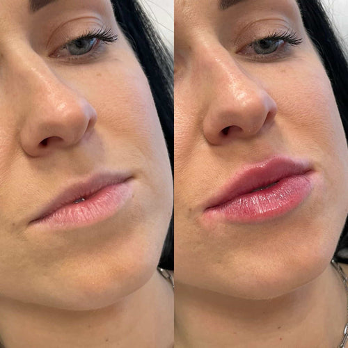
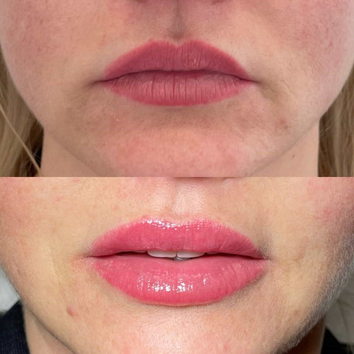
Dermal fillers have become increasingly popular in recent years, offering a non-surgical approach to enhance facial features and restore youthful contours. These injectable gels, typically made from hyaluronic acid, are carefully placed beneath the skin to add volume, smooth wrinkles, and create a more balanced and harmonious appearance.
What Are Dermal Fillers?
Hyaluronic acid is a naturally occurring substance in the body that helps maintain skin hydration and elasticity. Dermal fillers leverage this property by injecting a gel-like form of hyaluronic acid into specific areas of the face.
This added volume can plump up lips, reduce the appearance of fine lines and wrinkles, and redefine facial contours such as cheekbones or jawlines.
The results from dermal filler injections are typically visible immediately and can last anywhere from several months to over a year, depending on the type of filler used and individual factors like metabolism.
Types of Dermal Fillers
Dermal fillers have become increasingly popular in recent years, offering a non-surgical approach to enhance facial features and restore youthful contours. These injectable gels, typically made from hyaluronic acid, are carefully placed beneath the skin to add volume, smooth wrinkles, and create a more balanced and harmonious appearance.
Hyaluronic acid is a naturally occurring substance in the body that helps maintain skin hydration and elasticity. Dermal fillers leverage this property by injecting a gel-like form of hyaluronic acid into specific areas of the face.
This added volume can plump up lips, reduce the appearance of fine lines and wrinkles, and redefine facial contours such as cheekbones or jawlines.
The results from dermal filler injections are typically visible immediately and can last anywhere from several months to over a year, depending on the type of filler used and individual factors like metabolism.
There are various types of dermal fillers available, each with unique properties and applications:
Hyaluronic Acid Fillers: The most common type, these fillers provide temporary volume and hydration. They can be used for a variety of purposes, such as lip augmentation, wrinkle reduction, and cheek enhancement.
Poly-L-Lactic Acid (PLLA) Fillers: These fillers stimulate collagen production, gradually adding volume over time. Results typically last longer than hyaluronic acid fillers.
Calcium Hydroxylapatite Fillers: These fillers are biocompatible and designed to provide a more structured lift. They can be used to address deeper wrinkles and contouring needs.
How Dermal Fillers Work
Dermal fillers have become increasingly popular in recent years, offering a non-surgical approach to enhance facial features and restore youthful contours. These injectable gels, typically made from hyaluronic acid, are carefully placed beneath the skin to add volume, smooth wrinkles, and create a more balanced and harmonious appearance.
Hyaluronic acid is a naturally occurring substance in the body that helps maintain skin hydration and elasticity. Dermal fillers leverage this property by injecting a gel-like form of hyaluronic acid into specific areas of the face.
This added volume can plump up lips, reduce the appearance of fine lines and wrinkles, and redefine facial contours such as cheekbones or jawlines.
The results from dermal filler injections are typically visible immediately and can last anywhere from several months to over a year, depending on the type of filler used and individual factors like metabolism.
There are various types of dermal fillers available, each with unique properties and applications:
Hyaluronic Acid Fillers: The most common type, these fillers provide temporary volume and hydration. They can be used for a variety of purposes, such as lip augmentation, wrinkle reduction, and cheek enhancement.
Poly-L-Lactic Acid (PLLA) Fillers: These fillers stimulate collagen production, gradually adding volume over time. Results typically last longer than hyaluronic acid fillers.
Calcium Hydroxylapatite Fillers: These fillers are biocompatible and designed to provide a more structured lift. They can be used to address deeper wrinkles and contouring needs.
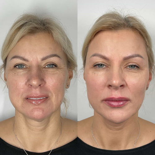
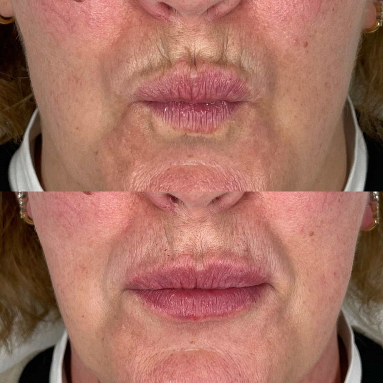
Facial Symmetry and Beauty Standards
Throughout history, symmetrical facial features have been associated with beauty standards across various cultures. A balanced and harmonious appearance is often perceived as attractive, leading to a desire to enhance or correct perceived asymmetries.
The Perception of Facial Harmony
Throughout history, symmetrical facial features have been associated with beauty standards across various cultures. A balanced and harmonious appearance is often perceived as attractive, leading to a desire to enhance or correct perceived asymmetries.
This perception of symmetry stems from evolutionary psychology, where it was linked to indicators of good health and genetic fitness. The human brain naturally seeks patterns and order, finding symmetrical faces more pleasing to the eye due to their inherent sense of balance.
While perfect symmetry is rare in nature, deviations that are subtle can often be imperceptible to the casual observer. However, more pronounced asymmetries can draw attention and potentially detract from perceived attractiveness.
Common Facial Asymmetries
Throughout history, symmetrical facial features have been associated with beauty standards across various cultures. A balanced and harmonious appearance is often perceived as attractive, leading to a desire to enhance or correct perceived asymmetries.
This perception of symmetry stems from evolutionary psychology, where it was linked to indicators of good health and genetic fitness. The human brain naturally seeks patterns and order, finding symmetrical faces more pleasing to the eye due to their inherent sense of balance.
While perfect symmetry is rare in nature, deviations that are subtle can often be imperceptible to the casual observer. However, more pronounced asymmetries can draw attention and potentially detract from perceived attractiveness.
Common facial asymmetries include differences in eyebrow height, nostril size, lip fullness, or ear positioning. These variations can occur naturally due to genetics or factors during development.
Using Fillers to Enhance Facial Symmetry
Throughout history, symmetrical facial features have been associated with beauty standards across various cultures. A balanced and harmonious appearance is often perceived as attractive, leading to a desire to enhance or correct perceived asymmetries.
This perception of symmetry stems from evolutionary psychology, where it was linked to indicators of good health and genetic fitness. The human brain naturally seeks patterns and order, finding symmetrical faces more pleasing to the eye due to their inherent sense of balance.
While perfect symmetry is rare in nature, deviations that are subtle can often be imperceptible to the casual observer. However, more pronounced asymmetries can draw attention and potentially detract from perceived attractiveness.
Addressing Specific Asymmetries
Throughout history, symmetrical facial features have been associated with beauty standards across various cultures. A balanced and harmonious appearance is often perceived as attractive, leading to a desire to enhance or correct perceived asymmetries.
This perception of symmetry stems from evolutionary psychology, where it was linked to indicators of good health and genetic fitness. The human brain naturally seeks patterns and order, finding symmetrical faces more pleasing to the eye due to their inherent sense of balance.
Contact Us
It’s Me and You Clinic – Anti-Wrinkle, Dermal Filler and Skincare Clinic, Kingston, Surrey
Kingston upon Thames , Survey, United KingdomKT2 6LX
While perfect symmetry is rare in nature, deviations that are subtle can often be imperceptible to the casual observer. However, more pronounced asymmetries can draw attention and potentially detract from perceived attractiveness.
Common facial asymmetries include differences in eyebrow height, nostril size, lip fullness, or ear positioning. These variations can occur naturally due to genetics or factors during development.
Dermal fillers offer a non-surgical solution to address these asymmetries and create a more balanced appearance. By strategically injecting filler into areas that need augmentation or contouring, practitioners can subtly enhance facial features and minimize the appearance of imbalances.
For example, if one side of the face appears droopy due to volume loss in the cheekbones, dermal filler can be used to add volume and lift the cheek on the weaker side, restoring symmetry.
Similarly, fillers can address unevenness in lip fullness or create a more symmetrical smile by subtly plumping up areas that need enhancement.
It is important to note that achieving facial symmetry with fillers is a delicate process that requires expertise and careful assessment. A qualified injector will consider the individual’s facial structure, anatomy, and desired outcome to develop a personalized treatment plan.
Creating Balance Through Subtle Adjustments
Dermal fillers have become increasingly popular in recent years, offering a non-surgical approach to enhance facial features and restore youthful contours. These injectable gels, typically made from hyaluronic acid, are carefully placed beneath the skin to add volume, smooth wrinkles, and create a more balanced and harmonious appearance.
Hyaluronic acid is a naturally occurring substance in the body that helps maintain skin hydration and elasticity. Dermal fillers leverage this property by injecting a gel-like form of hyaluronic acid into specific areas of the face.
This added volume can plump up lips, reduce the appearance of fine lines and wrinkles, and redefine facial contours such as cheekbones or jawlines.
The results from dermal filler injections are typically visible immediately and can last anywhere from several months to over a year, depending on the type of filler used and individual factors like metabolism.
Throughout history, symmetrical facial features have been associated with beauty standards across various cultures. A balanced and harmonious appearance is often perceived as attractive, leading to a desire to enhance or correct perceived asymmetries.
This perception of symmetry stems from evolutionary psychology, where it was linked to indicators of good health and genetic fitness. The human brain naturally seeks patterns and order, finding symmetrical faces more pleasing to the eye due to their inherent sense of balance.
While perfect symmetry is rare in nature, deviations that are subtle can often be imperceptible to the casual observer. However, more pronounced asymmetries can draw attention and potentially detract from perceived attractiveness.
Common facial asymmetries include differences in eyebrow height, nostril size, lip fullness, or ear positioning. These variations can occur naturally due to genetics or factors during development.
Dermal fillers offer a non-surgical solution to address these asymmetries and create a more balanced appearance. By strategically injecting filler into areas that need augmentation or contouring, practitioners can subtly enhance facial features and minimize the appearance of imbalances.
For example, if one side of the face appears droopy due to volume loss in the cheekbones, dermal filler can be used to add volume and lift the cheek on the weaker side, restoring symmetry.
Similarly, fillers can address unevenness in lip fullness or create a more symmetrical smile by subtly plumping up areas that need enhancement.
It is important to note that achieving facial symmetry with fillers is a delicate process that requires expertise and careful assessment. A qualified injector will consider the individual’s facial structure, anatomy, and desired outcome to develop a personalized treatment plan.
Considerations for Treatment
Dermal fillers have become increasingly popular in recent years for their ability to subtly enhance facial features and restore balance to the face. Achieving facial symmetry is often desired as it is perceived as more aesthetically pleasing.
Consultation and Assessment
When considering dermal fillers for facial symmetry enhancement, several crucial factors must be taken into account:
* **Consultation and Assessment:** A thorough consultation with a qualified injector is essential. This involves a detailed discussion of the patient’s goals, medical history, and any existing concerns. The injector will assess the patient’s facial anatomy, identify areas needing correction, and determine the most appropriate type and volume of filler to achieve the desired outcome.
* **Treatment Plan:** Based on the assessment, the injector will develop a personalized treatment plan outlining the specific injection points, techniques, and expected results. It is crucial for patients to understand the procedure, potential risks and benefits, and realistic expectations before making a decision.
* **Experience and Qualifications:** Choosing an experienced and qualified injector is paramount. Look for a practitioner with extensive training and experience in facial aesthetics, particularly in using dermal fillers for symmetry correction.
* **Type of Filler:** Different types of dermal fillers have varying properties and durations of effect. The injector will select the most suitable filler based on the individual’s needs and desired outcome.
* **Realistic Expectations:** It is important to have realistic expectations about what can be achieved with dermal fillers. While they can significantly enhance facial symmetry, they cannot completely correct severe asymmetries or achieve perfect symmetry.
Open communication between the patient and injector throughout the process is vital for ensuring a safe and successful outcome.
Choosing the Right Filler Type
When choosing the right dermal filler type, several factors should be considered:
Desired Outcome:
What specific areas need correction? Are you looking to add volume, smooth wrinkles, or define contours?
Duration of Effect:
Some fillers last longer than others. Hyaluronic acid fillers typically last 6-18 months, while poly-L-lactic acid (PLLA) fillers can last for up to two years.
Consistency and Spreadability:
The consistency of a filler determines how easily it spreads under the skin. Some fillers are thicker and more structured, ideal for contouring, while others are thinner and more fluid, better suited for smoothing wrinkles.
Patient’s Skin Type and Concerns:
An experienced injector will consider factors like skin elasticity, thickness, and any allergies or sensitivities when selecting the most appropriate filler type.
Treatment Procedure and Risks
Before undergoing dermal filler treatment for facial symmetry, potential risks and complications should be carefully considered:
Common Side Effects:
Most side effects from dermal fillers are mild and temporary, typically resolving within a few days to weeks. These can include bruising, swelling, redness, tenderness, itching, or small bumps at the injection site.
Serious Complications (Less Common):
While rare, some more serious complications can occur, such as:
Allergic Reactions:
Although uncommon, allergic reactions to the filler ingredients can happen.
Infection:
Any injection procedure carries a risk of infection if proper sterilization techniques are not followed.
Vascular Occlusion:
This occurs when a blood vessel is blocked by the filler, potentially leading to tissue damage or necrosis (death). It is crucial to avoid injecting filler into areas close to major blood vessels.
Asymmetry: **
Even with experienced injectors, achieving perfect symmetry can be challenging. There’s always a risk of one side looking different from the other.
Granuloma Formation: **
This is a small lump that forms as the body reacts to the filler material.
It is essential to consult with a qualified and experienced injector who can assess your individual needs, discuss potential risks, and ensure safe and effective treatment.
Choosing an accredited clinic or practice with appropriate licensing and sterile procedures further minimizes the risk of complications.
Long-Term Results and Maintenance
Dermal fillers offer a promising approach to enhance facial symmetry, but it’s crucial to understand that maintaining the results requires ongoing care and attention.
Replenishment Treatments:
As dermal fillers are temporary, repeat treatments are necessary to maintain the desired aesthetic outcome. The frequency of these touch-up appointments varies depending on the type of filler used, individual metabolism, and lifestyle factors. Typically, follow-up sessions are scheduled every 6 to 18 months.
Lifestyle Factors:
Certain habits can impact the longevity of dermal filler results. Protecting your skin from sun exposure through the use of sunscreen is essential as UV rays can break down hyaluronic acid, leading to premature filler degradation.
Smoking and excessive alcohol consumption can also affect collagen production and contribute to faster filler breakdown.
Maintaining a healthy lifestyle with a balanced diet, adequate hydration, and sufficient sleep supports overall skin health and helps preserve the long-term effects of dermal fillers.
Cheek fillers are a popular cosmetic procedure used to enhance the volume and definition of the cheeks.
They involve injecting dermal fillers, which are gel-like substances made from hyaluronic acid, into specific areas of the cheekbones.
Hyaluronic acid is naturally found in the body and helps to hydrate and plump the skin.
Dermal fillers can add volume to sunken cheeks, restore lost youthful fullness, and create a more sculpted appearance.
Here’s a breakdown of key information about cheek fillers:
* **What They Are:** Cheek fillers are injectable substances made from hyaluronic acid. Hyaluronic acid is naturally found in the body, attracting and holding water to keep skin hydrated and plump.
* **How They Work:** The filler is injected into specific areas of the cheeks by a qualified medical professional.
The gel-like consistency adds volume to enhance cheekbones, creating a more youthful and defined contour.
* **Types of Fillers:** There are different types of hyaluronic acid fillers available. Some popular brands include Juvederm Voluma XC, Restylane Lyft, and Radiesse. Each type may have slightly different properties and longevity.
* **Procedure:** The cheek filler procedure is typically quick and minimally invasive. It’s performed in a doctor’s office or clinic under local anesthesia. The process involves injecting the filler into the desired areas using a fine needle.
Aftercare Instructions: Following your treatment, you may experience some mild swelling, redness, or bruising. Your practitioner will provide specific instructions on how to care for the treated area.
Results typically last for several months, with touch-up appointments needed to maintain the desired fullness.
Types of Cheek Filler Material
Cheek fillers are a popular non-surgical cosmetic procedure used to enhance the volume and definition of the cheeks. They can help create a more youthful, sculpted appearance by adding fullness to sunken or hollow areas.
There are several types of cheek filler materials available, each with its own unique properties and benefits:
Hyaluronic Acid (HA) Fillers: These are the most common type of cheek fillers. HA is a naturally occurring substance in the body that helps to retain moisture and provide volume.
Contact Us
It’s Me and You Clinic – Anti-Wrinkle, Dermal Filler and Skincare Clinic, Kingston, Surrey
Kingston upon Thames, Surrey, United KingdomKT2 6LX
HA fillers are temporary, typically lasting 6-18 months, and can be dissolved with an enzyme called hyaluronidase if needed.
Popular brands of HA fillers include Juvederm, Restylane, and Belotero.
Poly-L-lactic Acid (PLLA) Fillers: PLLA fillers are made from biodegradable synthetic polymers.
Unlike HA fillers, they stimulate the body’s own collagen production, leading to longer-lasting results that can last up to 2 years or more.
A popular brand of PLLA filler is Sculptra Aesthetic.
Calcium Hydroxylapatite (CaHa) Fillers: CaHa fillers are another type of permanent filler. They are made from a synthetic material that mimics the body’s natural bone mineral structure.
CaHa fillers provide immediate volume and can last for several years. Radiesse is a common brand of CaHa filler.
Choosing the right cheek filler depends on individual needs, desired outcomes, and budget.
The Procedure
Cheek fillers are a popular non-surgical cosmetic procedure that can add volume, definition, and lift to the cheeks.
Understanding cheek fillers involves knowing the different types available, how they work, the procedure itself, potential risks and benefits, and what to expect during recovery.
The most common type, HA is a naturally occurring substance in the body that attracts and holds water, providing hydration and volume.
Popular brands include Juvederm Voluma XC and Restylane Lyft.
Calcium Hydroxylapatite (CaHA):
A biocompatible material that stimulates collagen production, leading to long-lasting results.
Radiesse is a well-known CaHA filler.
Poly-L-lactic Acid (PLLA):
Stimulates collagen production gradually over time, resulting in volumization that develops over several weeks or months.
Sculptra Aesthetic is a popular PLLA filler.
The Procedure:
Consultation:
A consultation with a qualified and experienced injector is essential. The injector will assess your facial structure, discuss your goals, and determine the best type of filler and amount needed.
Numbing Cream:
To minimize discomfort, a topical anesthetic cream will be applied to the injection area.
Injections:
Using a fine needle, the filler is injected into specific points on the cheeks. The injector may use techniques such as “fanning” or “sandwiching” to distribute the filler evenly and create a natural-looking result.
Ice Pack:
After the injections, an ice pack may be applied to reduce any swelling or bruising.
Recovery:
Downtime is minimal with cheek fillers. Some mild redness, swelling, and tenderness are common for a few days but usually subside within a week.
Avoid strenuous activity, excessive sun exposure, and touching the treated area immediately after injections.
Risks and Benefits:
Benefits:
Enhance cheek volume and definition.
Lift the cheeks and create a more youthful appearance.
Minimally invasive procedure with short recovery time.
Relatively affordable compared to surgery.
Risks:
Bruising, swelling, redness, and tenderness at the injection site.
Asymmetry or lumps if filler is not evenly distributed.
It’s crucial to consult with a board-certified dermatologist or plastic surgeon who specializes in fillers. They can evaluate your individual needs and determine if cheek fillers are right for you.
Benefits and Potential Drawbacks
Enhanced Facial Structure
Cheek fillers can be a popular choice for individuals looking to enhance their facial structure, but it’s essential to weigh the potential benefits against the possible drawbacks.
Benefits:
Enhanced Facial Volume and Definition: Cheek fillers can add volume to cheeks that have lost fullness due to aging or genetics, creating a more youthful and sculpted appearance. This can help define cheekbones, create a more prominent jawline, and improve facial symmetry.
Non-Surgical Solution: Unlike surgical procedures, cheek fillers are minimally invasive and don’t require incisions or anesthesia. They can be administered in a quick office visit with minimal downtime.
Reversible Results: Cheek filler injections are temporary, typically lasting 6 to 18 months depending on the type of filler used and individual factors. This allows for adjustments to be made as desired.
Natural-Looking Enhancement: When administered by a skilled injector, cheek fillers can create subtle, natural-looking enhancements that enhance rather than drastically alter facial features.
Potential Drawbacks:
Temporary Results: As mentioned, the effects of cheek fillers are not permanent and require repeat injections to maintain desired results. This can add to the overall cost of the procedure.
Risk of Side Effects: Like any medical procedure, cheek filler injections carry a risk of side effects, which can include bruising, swelling, redness, pain, and in rare cases, infection or allergic reaction.
Uneven Results: If the injections are not administered properly, it can result in uneven or unnatural-looking results. It’s crucial to choose an experienced and qualified injector.
Cost:** Cheek filler treatments can be expensive, particularly if multiple sessions are needed to achieve desired results.
Before making a decision about cheek fillers, it’s essential to consult with a board-certified plastic surgeon or dermatologist to discuss your goals, medical history, and potential risks and benefits.
Address Volume Loss
Cheek fillers are an increasingly popular cosmetic procedure that can provide a number of benefits, but also come with potential drawbacks.
One of the primary benefits of cheek fillers is their ability to enhance facial contours and volume. As we age, our cheeks naturally lose volume, leading to sagging skin and a more gaunt appearance. Fillers can plump up the cheeks, restoring lost definition and creating a more youthful look.
Fillers can also improve facial symmetry and balance. If one cheek is noticeably larger or smaller than the other, fillers can be used to even things out and create a more harmonious look. They can also enhance the appearance of smile lines and nasolabial folds by adding volume and smoothing out wrinkles.
The results of cheek filler injections are typically immediate and can last anywhere from six months to two years, depending on the type of filler used and individual factors.
However, there are also potential drawbacks to consider. Like any medical procedure, cheek fillers carry some risk of side effects, which can include bruising, swelling, redness, pain, and infection.
There is also a risk of complications such as vascular occlusion (blockage of a blood vessel) or filler migration. In rare cases, these complications can be serious.
Additionally, the results of cheek fillers are not permanent. As the filler gradually breaks down over time, the cheeks will lose volume and the effects will fade. This means that touch-up appointments will be necessary to maintain the desired look.
Another potential drawback is the cost of cheek fillers, which can vary depending on the type of filler used and the amount injected.
It’s important to consult with a qualified and experienced plastic surgeon or dermatologist to discuss your goals and expectations for cheek fillers. They can assess your individual suitability for the procedure, explain the potential risks and benefits in detail, and help you make an informed decision about whether cheek fillers are right for you.
Risks and Complications
Cheek fillers offer a non-surgical way to enhance facial volume and contours, but like any cosmetic procedure, they come with both benefits and potential drawbacks.
**Benefits:**
Facial Rejuvenation: Fillers can restore lost volume in the cheeks, reducing the appearance of wrinkles and creating a more youthful, lifted look.
Enhanced Facial Symmetry: Fillers can be used to balance out asymmetrical features, such as uneven cheekbones.
Improved Facial Definition: By adding volume to the cheeks, fillers can create a more defined and sculpted appearance.
Non-Surgical Procedure: Unlike surgery, filler injections are minimally invasive, requiring no incisions or lengthy recovery time.
Quick Treatment Time: Filler injections typically take only 15-30 minutes to perform.
**Potential Drawbacks and Complications:**
Temporary Results: The effects of cheek fillers are not permanent, typically lasting 6-18 months, depending on the type of filler used and individual factors.
Injection Site Reactions: Some common side effects include redness, swelling, bruising, and tenderness at the injection sites. These usually subside within a few days.
Asymmetry or Lumpiness: If fillers are not injected evenly, it can lead to asymmetry or an unnatural lumpiness in the cheeks.
Allergic Reactions: While rare, some individuals may experience allergic reactions to the filler material.
Infection: There is a small risk of infection at the injection site.
**Risks:**
Vascular Occlusion: Injecting filler into or near a blood vessel can obstruct blood flow, leading to tissue damage.
Nerve Damage: Although rare, improper injection techniques can damage nerves, potentially causing numbness or weakness in the face.
It’s important to consult with a qualified and experienced injector to discuss your desired outcome and any potential risks or complications. They will assess your individual needs and medical history to determine if cheek fillers are right for you.
Considerations Before Getting Cheek Fillers
Consultation with a Qualified Provider
Cheek fillers are a popular cosmetic procedure that can enhance facial contours and restore lost volume. However, it’s crucial to carefully consider several factors before deciding if cheek fillers are right for you.
Firstly, understand your goals and expectations. What specific changes are you hoping to achieve? Do you want to add definition, lift sagging cheeks, or correct asymmetry? Clearly defining your desired outcome will help guide your decision and communication with your provider.
Next, research different types of fillers available. Hyaluronic acid fillers are commonly used for cheek augmentation due to their versatility and ability to naturally integrate with the body. They come in various viscosities, allowing for customized results. Some fillers offer longer-lasting effects than others. Discuss your options with a qualified provider to determine the best fit for your needs.
Consider your budget as filler treatments can vary significantly in cost depending on factors like the amount of product used, the provider’s experience, and geographic location. Factor in potential follow-up appointments and touch-ups needed to maintain your desired results.
A thorough consultation with a qualified medical professional is essential before undergoing any cosmetic procedure. Look for a board-certified dermatologist, plastic surgeon, or otolaryngologist (ENT doctor) with extensive experience in facial fillers.
During the consultation, be prepared to discuss your medical history, allergies, medications, and any previous cosmetic procedures. Share your goals and desired outcome with honesty and openness. A qualified provider will assess your anatomy, skin type, and individual needs to determine if cheek fillers are appropriate for you.
The consultation is also an opportunity to ask questions about the procedure, potential risks and side effects, aftercare instructions, and expected results. Don’t hesitate to inquire about before-and-after photos of previous patients to get a sense of the provider’s artistry and expertise.
Choosing a reputable provider who prioritizes patient safety and provides personalized care is paramount for a successful and satisfying outcome with cheek fillers.
Realistic Expectations
Cheek fillers are a popular cosmetic procedure that can enhance facial volume and contour, creating a more youthful and defined appearance. However, it’s essential to carefully consider several factors before undergoing this treatment to ensure it aligns with your expectations and goals.
One of the most important considerations is your individual anatomy and facial structure. Cheek fillers work best on individuals who have naturally hollow or flat cheeks, as they can add volume and projection. If your cheekbones are already prominent, fillers may not be necessary or may lead to an over-filled appearance.
Realistic expectations are crucial for a successful outcome. Fillers can enhance your natural features but cannot completely transform your face. It’s important to discuss your desired results with a qualified and experienced injector who can assess your unique facial structure and provide realistic recommendations.
Another factor to consider is the type of filler material used. Different fillers have varying properties, such as longevity, consistency, and lift. Hyaluronic acid fillers are commonly used for cheek augmentation due to their natural-looking results and ability to be reversed if necessary.
The cost of cheek fillers can vary widely depending on factors such as the amount of filler required, the injector’s experience, and geographical location. It’s essential to obtain a clear understanding of all costs involved before undergoing the procedure.
Potential side effects, although usually mild and temporary, should be discussed with your injector. These may include bruising, swelling, redness, and tenderness at the injection site.
Finally, it’s important to choose a qualified and experienced injector who is licensed and board-certified in a relevant medical specialty. A reputable injector will prioritize patient safety and provide personalized care throughout the process.
Long-Term Costs
Cheek fillers can be a popular cosmetic procedure to enhance facial contours and add volume, but it’s essential to carefully consider various factors before making a decision.
Here are some key considerations:
Goals and Expectations: Clearly define your desired outcome. What do you hope to achieve with cheek fillers? Do you want a subtle lift, more definition, or a dramatic change? Realistic expectations are crucial for achieving satisfaction.
Consultation with a Qualified Professional: It’s vital to consult with a board-certified plastic surgeon or dermatologist experienced in facial filler injections. They can assess your facial structure, skin type, and overall health to determine if fillers are suitable for you and discuss the potential risks and benefits.
Type of Filler: Different types of fillers exist, each with unique properties and longevity. Hyaluronic acid fillers, such as Juvederm or Restylane, are commonly used for cheek augmentation due to their ability to attract and hold water, providing natural-looking results.
Cost: Cheek filler injections can range in price depending on the amount of filler used, the injector’s experience, and geographical location. Discuss the cost upfront with your chosen provider to understand the total expenses involved.
Long-Term Costs:** While cheek fillers typically last 6 to 18 months, it’s important to consider the long-term costs of maintenance. Repeat injections will be needed to maintain the desired results.
Budget for these touch-up appointments over time.
Potential Side Effects: Like any medical procedure, cheek filler injections carry potential side effects. These can include bruising, swelling, redness, tenderness, and in rare cases, infections or allergic reactions. Discuss these risks with your provider beforehand.
Aftercare Instructions: Carefully follow your provider’s aftercare instructions to minimize the risk of complications and ensure optimal healing.
By thoughtfully considering these factors, you can make an informed decision about whether cheek fillers are right for you.
Many factors influence how long it takes for lip fillers to dissolve naturally. The primary factor is the *type* of filler used.
**Hyaluronic Acid (HA)** fillers are the most popular type, and they dissolve gradually over time due to the body’s natural breakdown of hyaluronic acid. Dissolution typically occurs within six months to two years, depending on several factors.
Dermal Fillers Composed of Poly-L-lactic Acid (PLLA) are different. PLLA is a synthetic material that stimulates collagen production. It gradually dissolves over time as the body reabsorbs it, but this process takes much longer than with HA fillers, often lasting for several years.
Other factors influencing dissolution time include:
* **Filler Injection Technique:** The deeper and more evenly the filler is injected, the slower it may dissolve as it has less direct contact with surrounding tissues.
* **Metabolic Rate:** Individuals with faster metabolisms tend to metabolize fillers quicker.
* **Age:** As we age, collagen production decreases, which can affect how long fillers last.
* **Sun Exposure:** Excessive sun exposure can break down hyaluronic acid fillers, causing them to dissolve faster.
Individual Metabolism
Dissolution time for lip fillers depends on a variety of factors, making it impossible to give a definitive timeframe.
Here’s a breakdown of key influences:
* **Type of filler:** Different dermal fillers are composed of various substances and designed to last different durations.
Hyaluronic acid (HA) fillers, the most common type, tend to dissolve within 6 months to 2 years.
Permanent fillers, containing materials like silicone or collagen, are not naturally dissolvable and require surgical removal.
* **Individual metabolism:** This plays a significant role. People with faster metabolisms may experience quicker filler breakdown.
*
Injection location:
Areas with higher blood flow, like the lips, often see faster dissolution compared to areas with slower circulation.
Lip fillers are typically absorbed more quickly due to the increased blood supply in this area.
* **Lifestyle factors:**
Smoking can accelerate filler breakdown by reducing blood flow and oxygenation.
Excessive sun exposure can break down collagen, potentially influencing filler longevity.
*
Age:
As we age, our skin’s collagen production naturally declines, which may affect how long fillers remain visible.
It’s crucial to remember that these are general guidelines. Consult with a qualified medical professional for personalized information regarding your specific situation and expected dissolution time for your lip filler.
Location of Injection
Dissolution time for lip fillers depends on several factors, primarily the type of filler used and individual body chemistry.
Here are some key factors affecting dissolution time:
Filler Type: Hyaluronic acid (HA) fillers, the most common type, naturally degrade over time as the body reabsorbs them. Different HA fillers have varying molecular weights and cross-linking densities, influencing their lifespan. Generally, smaller molecules and less cross-linking lead to faster dissolution.
Body Metabolism: Individual metabolism rates significantly affect filler breakdown. People with faster metabolisms tend to see fillers dissolve quicker.
Injection Location: Fillers injected into areas with higher blood flow, like the lips, generally dissolve faster than those in areas with lower blood flow. Blood circulation helps transport enzymes that break down the filler.
Sun Exposure: Excessive sun exposure can degrade hyaluronic acid fillers, leading to faster dissolution.
Lifestyle Factors: Smoking and alcohol consumption may affect collagen production and overall tissue health, potentially influencing filler longevity.
Contact Us
It’s Me and You Clinic – Anti-Wrinkle, Dermal Filler and Skincare Clinic, Kingston, Surrey
Kingston upon Thames, Surrey, United KingdomKT2 6LX
While it’s impossible to give a precise timeframe for natural filler dissolution, HA fillers typically last anywhere from 6 months to 2 years, depending on the factors listed above.
For specific information regarding the expected lifespan of your particular lip filler type, consult with your injector.
Estimating the exact timeline for *lip filler* dissolution is challenging as it varies significantly between individuals and depends on several factors.
Here’s a general range you can expect:
Initial Reduction: Within the first few weeks, most people notice a gradual reduction in volume as the hyaluronic acid filler begins to be broken down by the body.
Significant Dissolution: Over 3-6 months, you’ll likely see a noticeable decrease in filler, and the lips will start to regain their natural shape more prominently.
Full Dissolution (Approximation): For most individuals, full dissolution of hyaluronic acid fillers takes around 6-18 months. However, it’s essential to remember this is an estimate and not a definitive timeline.
Factors influencing the dissolution rate include:
Type of filler used:** Different brands and formulations have varying compositions that affect how quickly they are metabolized.
Amount of filler injected:** Larger injections naturally take longer to dissolve than smaller ones.
Individual metabolism:** Everyone’s body processes substances differently. Some individuals may break down fillers faster than others.
Lifestyle factors:** Things like hydration, diet, and smoking can potentially influence filler breakdown.
If you have any concerns about your lip filler dissolving timeline or want a more precise estimate, it’s best to consult with your injector. They can assess your specific situation and provide tailored advice.
Potential Variation
Estimated dissolution timelines for lip fillers vary widely depending on several factors, primarily the specific type of filler used.
Hyaluronic acid (HA) fillers, the most common type, are typically broken down by the body within 6-18 months.
The exact duration can be influenced by individual metabolism, injection technique, and product formulation. For example, some HA fillers may contain additives or crosslinking agents that affect their longevity.
Other types of fillers, like poly-L-lactic acid (PLLA), work differently. Instead of being directly broken down, they stimulate collagen production, resulting in a gradual volumizing effect that can last for 12-24 months or longer.
Individual variations are significant. Lifestyle factors like sun exposure and smoking can accelerate filler breakdown, while healthy habits may prolong its effects.
It’s crucial to remember that these are just estimations, and the actual dissolution timeline can vary considerably from person to person.
Regular follow-up appointments with a qualified injector allow for personalized assessments and adjustments to treatment plans based on individual progress.
Lip filler typically dissolves gradually over time due to the body’s natural process of breaking down foreign substances.
The rate of dissolution varies depending on several factors, including the type of filler used, individual metabolism, and lifestyle habits. Hyaluronic acid fillers, which are the most common type, tend to last longer than other types of fillers.
Here are some signs that your lip filler is dissolving or reducing in volume:
• Gradual loss of fullness: You may notice a subtle decrease in the plumpness and definition of your lips over time.
• Flattening of lip contours: The edges of your lips may appear softer and less defined as the filler dissolves.
• Visible lines and wrinkles: As the filler diminishes, existing fine lines or wrinkles around the mouth may become more prominent.
• Asymmetry: If you had filler injected asymmetrically, one lip may start to dissolve faster than the other, resulting in an imbalance.
It’s important to note that these changes typically occur gradually over several months. However, some individuals may experience a more rapid reduction in volume due to their unique body chemistry or factors such as excessive sun exposure or dehydration.
If you are concerned about the longevity of your lip filler or notice any unusual changes, it is best to consult with a qualified medical professional who can assess your individual situation and provide appropriate guidance.
Texture Changes
Dissolving lip filler isn’t always a straightforward process with a clear timeline. It depends on several factors, including the type of filler used, individual metabolism, and location of the injections.
However, certain signs can indicate that your lip filler is gradually dissolving. Keep in mind these are general observations and individual experiences may vary.
One prominent sign is a change in fullness. You might notice your lips becoming less plump and defined over time. This gradual loss of volume is the most noticeable indicator of the filler breaking down.
Texture changes can also be apparent. As the hyaluronic acid-based filler degrades, it may start to feel softer or more spread out. Your lips might lose their initial smooth, sculpted appearance and seem slightly uneven.
Another sign is a shift in shape. Initially, lip filler enhances volume and contours. As it dissolves, your natural lip shape may become more pronounced, potentially leading to a less defined cupid’s bow or smoother borders.
While these changes are subtle at first, they become more noticeable as the filler continues to break down.
It’s important to remember that everyone’s body processes filler differently, so there’s no fixed timeframe for complete dissolution.
Loss of Shape
Signs that lip filler may be dissolving include:
• Gradual reduction in lip fullness.
• A change in the shape of the lips, often becoming more subtle or losing definition.
• A softening of previously plumped areas, making lips appear less defined and fuller.
• Subtle puddling or unevenness in the treated area.
The rate at which lip filler dissolves naturally varies depending on several factors:
Type of filler used: Some fillers are designed to dissolve more quickly than others.
Individual metabolism: How quickly your body metabolizes hyaluronic acid (the main component of most fillers) influences the duration of results.
Lifestyle factors: Sun exposure, smoking, and dehydration can accelerate filler breakdown.
Treatment location: Fillers placed in areas with more movement, like the lips, tend to dissolve faster than those in static areas.
It’s important to note that these signs may also be caused by other factors, so consulting a qualified medical professional is crucial for accurate diagnosis and advice.
Kingston upon Thames, Surrey, United KingdomKT2 6LX
Lip filler longevity can vary greatly from person to person, even when using the same type and amount of filler. Several factors influence how long your lip fillers will last, including metabolism and body chemistry, as well as your lip movement and expression.
**Metabolism and Body Chemistry**
Enzymatic Degradation: The body naturally breaks down hyaluronic acid fillers (the most common type) through an enzyme called hyaluronidase. People who metabolize this enzyme more quickly may see their filler dissolve faster.
Individual Body Chemistry: Everyone’s body chemistry is unique. Some individuals may have a higher rate of collagen turnover or other factors that affect how the filler integrates and degrades.
Genetics:**
There is evidence suggesting that genetics can play a role in how long fillers last.
Lip Movement and Expression
**Frequency of Use:** If you frequently purse your lips, smile, or pucker, the filler will be subjected to more stress and may break down faster.
**Muscle Activity:** People who have naturally more active lip muscles may experience quicker degradation of fillers.
Other Factors Influencing Longevity
Filler Type: Some fillers are designed to last longer than others. Discuss your expectations and lifestyle with your injector to choose the most appropriate filler for you.
Injection Technique:** A skilled injector can maximize longevity by placing filler strategically and ensuring proper distribution.
**Aftercare: Following your injector’s instructions carefully regarding aftercare can help protect the filler and prolong its effects.
Lip filler longevity can vary greatly from person to person, and several factors contribute to this variation. Two key areas influencing how long fillers last are metabolism and body chemistry, along with muscle activity around the injection site.
Metabolism refers to the body’s process of breaking down substances for energy and repair. Individuals with faster metabolisms tend to metabolize hyaluronic acid fillers (the most common type) more quickly, leading to shorter-lasting results.
Body chemistry also plays a role. Factors like skin elasticity, hydration levels, and the presence of certain enzymes can influence how well fillers integrate and remain stable in the tissues.
Muscle activity is another significant factor. The lips are constantly in motion for speaking, eating, and smiling. Areas with more movement will naturally break down filler particles faster. People who purse their lips frequently or have a habit of smoking may experience shorter-lasting results.
Individual Healing and Immune Response
There are several factors that can influence how long lip filler lasts, and understanding these can help you manage your expectations and make informed decisions about your treatments.
One significant factor is **metabolism**. Everyone metabolizes substances differently. Some individuals have a faster metabolism, which means the body breaks down hyaluronic acid (the most common type of filler) more quickly.
**Body chemistry** plays a role as well. Factors like hydration levels, hormonal fluctuations, and even skin elasticity can affect how filler is absorbed and distributed.
Individual healing and immune response are also crucial. Your body’s natural reaction to foreign substances (like fillers) can vary significantly. Some people may have an exaggerated immune response, leading to faster breakdown of the filler.
Additionally, **lifestyle factors** like smoking, excessive sun exposure, and dehydration can all contribute to quicker filler degradation.
It’s important to remember that lip fillers are temporary and will eventually be absorbed by the body. The longevity can range from several months to a year or more depending on the individual and the factors mentioned above.
If you’re concerned about your lip filler not lasting as long as you’d like, discuss your concerns with a qualified aesthetic professional. They can help you understand your individual circumstances and recommend options for maximizing results.
Enhancing Your Lip Filler Results
Choosing the Right Filler
Lip filler results can vary greatly from person to person and depend on a multitude of factors, including individual metabolism, lip anatomy, lifestyle habits, and the type of filler used.
One reason why lip filler might not last as long for some individuals is due to their natural lip physiology. Thinner lips or those with naturally more mobile muscles may break down filler quicker.
Lifestyle choices also play a significant role. Smoking, excessive sun exposure, and dehydration can all accelerate the breakdown of hyaluronic acid fillers.
Choosing the right type of filler is crucial for achieving longer-lasting results. Hyaluronic acid (HA) fillers are a popular choice due to their versatility, natural appearance, and ability to be dissolved if necessary. Different HA fillers have varying degrees of viscosity and crosslinking, which affect how long they last.
A highly crosslinked filler will generally provide longer-lasting results compared to a less crosslinked one.
When considering lip filler, consult with an experienced and board-certified injector. They can assess your individual needs and recommend the best type of filler and treatment plan for you.
During your consultation, discuss your desired outcome, medical history, and any concerns you may have. A good injector will take the time to understand your goals and provide realistic expectations regarding the longevity of results.
Viscosity and Formulation
Lip fillers are a popular cosmetic procedure that can add volume, definition, and fullness to the lips. However, some individuals may find that their lip filler results don’t last as long as expected.
Several factors can contribute to shorter-lasting lip filler results, including metabolism, lifestyle choices, and product viscosity and formulation.
Viscosity refers to a gel’s thickness or resistance to flow. A thicker, more viscous filler will typically last longer than a thinner one because it takes more time for the body to break down.
The formulation of lip filler also plays a crucial role in longevity. Fillers are typically composed of hyaluronic acid (HA) cross-linked with BDDE (bis-dioxidoethylidene). The degree of crosslinking affects how long the filler lasts.
Higher crosslinking creates a more stable and durable gel that resists degradation, resulting in longer-lasting results.
Furthermore, fillers containing additional ingredients, such as stabilizers or antioxidants, can help improve their longevity by protecting against breakdown and premature degradation.
When considering lip filler treatments, it’s important to discuss your expectations with a qualified and experienced injector. They can assess your individual needs, skin type, and lifestyle factors to recommend the most suitable filler type and crosslinking level for optimal results and longevity.
Ultimately, achieving long-lasting lip filler results requires a combination of factors, including choosing the right product, proper injection techniques, and maintaining a healthy lifestyle.
Placement Technique
One of the most common reasons lip filler might not last as long for some individuals is improper placement technique.
Optimal Placement is crucial for both achieving a natural-looking result and maximizing longevity.
When filler is placed superficially, it’s more likely to be absorbed by the body sooner. Conversely, injecting too deep can lead to lumpiness or displacement, ultimately reducing the lifespan of the filler.
Experienced Injectors: A skilled injector understands the subtle anatomy of the lips and will precisely target different areas for optimal volume and shape. They consider factors like muscle movement and natural lip contours to ensure a balanced and long-lasting result.
Desired Look:** Different placement techniques achieve distinct aesthetic outcomes. For example, placing filler in the **border** of the lips can create a fuller pout, while injecting into the center enhances cupid’s bow definition.
Open communication with your injector is essential. Discuss your desired look and any concerns you have about the longevity of lip fillers.
They can assess your individual needs, skin type, and lifestyle to recommend a suitable filler type and placement technique that maximizes results and duration.
Aftercare Practices
Lip filler results can vary significantly from person to person and are influenced by a variety of factors, including metabolism, lifestyle choices, and individual skin characteristics.
While lip fillers are designed to provide long-lasting enhancement, there are certain aftercare practices that can help maximize their longevity and ensure optimal results.
Following Injection:
Immediately after your lip filler treatment, it’s crucial to follow your injector’s specific instructions. This typically includes avoiding strenuous activity, touching or rubbing the treated area, and consuming alcohol for at least 24 hours.
Applying ice packs wrapped in a thin cloth can help reduce swelling and discomfort during the initial recovery period.
Hydration and Nourishment:
Drinking plenty of water is essential to maintain hydration, which supports collagen production and skin health. A balanced diet rich in vitamins, especially vitamin C, can also contribute to plumpness and longevity.
Consider incorporating hyaluronic acid supplements into your routine, as hyaluronic acid is a key component of lip filler.
Sun Protection:
UV radiation from the sun can break down collagen and elastin, which contribute to skin volume. Protect your lips by wearing broad-spectrum sunscreen with an SPF of 30 or higher daily, even on cloudy days. Consider a lip balm with SPF for added protection.
Lip Care Products:
Avoid using harsh scrubs or exfoliants on the treated area as they can irritate and damage the skin. Opt for gentle cleansers and moisturizers specifically designed for sensitive skin.
Consider using a lip balm with hydrating ingredients such as shea butter, coconut oil, or hyaluronic acid to keep lips soft and supple.
Smoking and excessive alcohol consumption can accelerate collagen breakdown and negatively impact filler longevity. Limiting these habits can help maintain your results for a longer duration.
Remember that individual results may vary, and it’s essential to consult with a qualified and experienced injector for personalized advice and treatment recommendations.
Additional Tips for Maximizing Filler Duration
Maintaining Hydration
Maximizing the longevity of lip filler requires a multi-faceted approach that goes beyond simply getting the injections.
Here are some additional tips to help you make the most of your filler and keep your lips hydrated:
Pre-Treatment Preparation:**
• Choose the Right Provider: Selecting a qualified and experienced injector is crucial. Look for someone with expertise in facial anatomy, dermal fillers, and lip augmentation.
• Understand Your Skin Type: Different skin types metabolize filler at varying rates. Discuss your skin type and concerns with your injector to determine the best product and treatment plan.
Post-Treatment Care:**
• Follow Instructions Carefully: Adhere to all post-treatment instructions provided by your injector, including avoiding strenuous activity, sun exposure, and certain skincare products.
• Ice Packs for Swelling: Apply ice packs wrapped in a towel for 15-20 minutes at a time throughout the first 48 hours to minimize swelling and bruising.
• Gentle Cleansing: Wash your face twice daily with a mild, fragrance-free cleanser. Avoid harsh scrubbing or exfoliating.
• Avoid Touching and Picking: Resist the urge to touch or pick at the injected area, as this can increase the risk of infection and migration of the filler.
Long-Term Hydration Strategies:**
• Stay Hydrated: Drink plenty of water throughout the day to keep your skin plump and hydrated from within.
• Lip Balm Application: Apply a hydrating lip balm regularly, especially before bedtime. Look for balms with hyaluronic acid or other moisturizing ingredients.
• Avoid Dehydrating Habits: Limit caffeine and alcohol intake, as they can dehydrate the body and lips.
• Protect from Sun Damage: Wear sunscreen daily, even on cloudy days, to prevent premature aging and breakdown of collagen.
Additional Tips:**
• Consider Lip Augmentation Supplements: Discuss with your injector about supplements like hyaluronic acid or collagen peptides that may support lip volume.
• Regular Touch-Up Appointments: Schedule follow-up appointments with your injector as recommended to maintain the desired results and address any concerns.
Protecting from Sun Exposure Avoiding Smoking and Excessive Alcohol Consumption
While everyone’s metabolism and skin type differ, there are additional tips you can follow to potentially extend the duration of your lip filler results.
Sun Exposure: Just as with any part of your face, excessive sun exposure can break down hyaluronic acid fillers. Always wear a high-quality *broad-spectrum sunscreen* (SPF 30 or higher) daily on your lips, even on cloudy days. Consider using lip balms containing SPF for added protection.
Smoking: Nicotine in cigarettes constricts blood vessels, hindering the circulation of nutrients and oxygen to the injection site. This can contribute to faster filler breakdown. If you smoke, quitting is one of the best things you can do for your overall health and the longevity of your fillers.
Excessive Alcohol Consumption: While moderate alcohol intake might not be drastically detrimental, excessive drinking can dehydrate your body, including the delicate skin on your lips. Dehydration can lead to faster filler breakdown and a less plump appearance.
It’s important to have realistic expectations about filler longevity. Factors like individual metabolism, lifestyle habits, and the specific type of filler used will all influence how long it lasts.
A ponytail facelift, also known as a short-scar facelift or minimally invasive facelift, is a cosmetic surgery procedure designed to rejuvenate the lower face and neck by subtly lifting and tightening sagging tissues.
This technique focuses on addressing signs of aging such as jowls, wrinkles, and loose skin around the jawline and neck.
It’s considered less invasive than a traditional facelift because it involves smaller incisions typically hidden within the hairline or behind the ears.
The procedure is performed under general anesthesia and can take approximately 2 to 3 hours. During surgery, the surgeon makes these incisions:
Behind the ear
Within the hairline
These incisions allow access to the underlying facial tissues.
The surgeon then:
Lifts and tightens the sagging skin and muscles of the face and neck
Excess skin is trimmed away
The incisions are closed with sutures.
Recovery from a ponytail facelift is typically shorter than a traditional facelift, with most patients returning to normal activities within a week or two.
However, it’s essential to follow the surgeon’s postoperative instructions carefully to minimize scarring and ensure optimal healing.
Results of a ponytail facelift can last for several years, depending on individual factors such as genetics, skin elasticity, and lifestyle habits.
Recovery
A ponytail facelift, also known as a *limited incision facelift*, targets sagging skin and fat in the lower face and neck. It focuses primarily on restoring the area around the jawline and neckline, often leaving the forehead and upper eyelids untouched.
The procedure typically involves incisions that follow natural contours, such as behind the ears or within the hairline. This allows for a more subtle scar placement compared to traditional facelifts.
During surgery, the surgeon will lift and reposition underlying facial tissues, removing excess skin and fat as needed. They may also address *platysmal bands*, which are muscle cords that can contribute to a loose or “turkey neck” appearance.
The recovery from a ponytail facelift is generally considered less invasive than a full facelift. Most patients experience minimal swelling and bruising, and can return to work within a few days to a week.
Full recovery may take several weeks, during which time it’s important to follow post-operative instructions carefully. This includes avoiding strenuous activity, keeping the incision area clean, and wearing compression garments as instructed.
Patients can expect gradual improvement in their appearance over time as swelling subsides and tissues heal. Final results are usually visible within 3 to 6 months.
Benefits
Addressing Sagging Skin
Contact Us
It’s Me and You Clinic – Anti-Wrinkle, Dermal Filler and Skincare Clinic, Kingston, Surrey
Kingston upon Thames, Surrey, United KingdomKT2 6LX
A ponytail facelift, also known as a mini facelift or a limited incision facelift, is a less invasive alternative to traditional facelifts. It focuses on tightening the skin and tissues along the lower third of the face, addressing areas like sagging jowls, loose skin under the chin (turkey neck), and fine lines around the mouth.
This procedure utilizes shorter incisions compared to traditional facelifts, typically hidden within the natural creases behind the ear and along the hairline. The surgeon lifts and tightens the underlying facial tissues, removing excess skin for a smoother and more contoured appearance.
The benefits of a ponytail facelift include:
Improved Facial Contour: Tightening loose skin and repositioning sagging tissues results in a more defined jawline, reduced jowls, and an overall lifted appearance.
Minimized Neck Aging: The procedure addresses “turkey neck” by tightening the skin and muscles in the lower neckline, creating a smoother transition between the face and neck.
Smoother Forehead and Eyebrows: While primarily focused on the lower face, a ponytail facelift can often lift sagging eyebrows, further enhancing facial rejuvenation.
Reduced Fine Lines and Wrinkles: By tightening the skin, fine lines around the mouth and lower jaw are minimized, resulting in a more youthful complexion.
Compared to traditional facelifts, ponytail facelift offers several advantages:
Book Your Dermal Filler Consultation with Dr. Laura Geige Today
1. **Less Invasive:** Shorter incisions and less extensive dissection translate into reduced trauma to surrounding tissues.
2. **Shorter Recovery Time:** Patients typically experience less bruising, swelling, and discomfort, leading to a faster return to normal activities.
3. **Minimal Scarring:** The incisions are strategically placed in natural folds, resulting in discreet scars that blend well with the hairline and behind the ears.
4. **Natural-Looking Results:** By focusing on subtle rejuvenation, ponytail facelifts maintain a natural appearance without creating an overly taut or pulled look.
Minimally Invasive Approach
A minimally invasive approach to a ponytail facelift, often referred to as a “mini lift” or “limited incision facelift,” offers several advantages over traditional facelift procedures.
Here are some key benefits:
Less Invasive: The incisions are smaller and strategically placed in natural hairline creases, minimizing scarring.
Shorter Recovery Time: Expect a quicker return to normal activities compared to traditional facelifts. Swelling and bruising typically subside faster.
Reduced Discomfort: Minimally invasive techniques often involve local anesthesia with sedation, leading to less overall discomfort during and after the procedure.
Natural-Looking Results: A mini facelift focuses on rejuvenating specific areas, such as the lower face and neck, while preserving a more natural appearance.
However, it’s important to note that the results of a minimally invasive ponytail facelift may not be as dramatic or long-lasting as those achieved with a traditional facelift. It’s best suited for individuals with mild to moderate signs of aging and realistic expectations.
During a consultation with a qualified plastic surgeon, you can discuss your specific concerns and determine if a mini facelift is the right approach for you.
Considerations
Consultation with a Surgeon
A ponytail facelift, also known as a mini facelift or short scar facelift, is a less invasive surgical procedure compared to a traditional facelift. It focuses primarily on addressing sagging skin and tissues in the lower face and neck, particularly along the jawline and cheek area.
When considering a ponytail facelift, it’s crucial to consult with a board-certified plastic surgeon. This consultation provides an opportunity to discuss your goals, concerns, and expectations for the procedure. The surgeon will assess your facial anatomy, skin elasticity, and overall health to determine if you’re a suitable candidate.
During the consultation, it’s essential to ask questions to ensure you fully understand the procedure, its potential benefits, risks, and recovery process.
Here are some key considerations for discussion with your surgeon:
• **Your Goals:** Be clear about what you hope to achieve with the ponytail facelift. Do you want to tighten the skin around your jawline? Reduce neck bands? Improve facial contour?
• **Your Expectations:** Understand that a ponytail facelift is not a complete rejuvenation. It primarily addresses moderate sagging in the lower face and neck. Discussing realistic outcomes based on your age, skin condition, and lifestyle is important.
• **Surgical Technique:** Inquire about the surgeon’s experience with ponytail facelifts, the incision placement (usually hidden behind the ears and within the hairline), and the type of sutures used.
• **Recovery Process:** Understand the expected downtime, including bruising, swelling, and limitations on activity. Discuss pain management options and any potential side effects.
• **Anesthesia:** Clarify what type of anesthesia will be used (local or general) and discuss any relevant medical history or allergies you have.
• **Cost and Financing:** Inquire about the total cost of the procedure, including surgeon fees, facility charges, anesthesia costs, and any potential follow-up visits. Explore financing options if needed.
• **Aftercare Instructions:** Understand what steps you need to take post-surgery for optimal healing, including wound care, medication, and activity restrictions.
• **Follow-Up Appointments:** Schedule a follow-up appointment with the surgeon to monitor your recovery progress and address any questions or concerns.
Choosing a qualified and experienced plastic surgeon is paramount. Ensure they have board certification from the American Board of Plastic Surgery (ABPS). Request before-and-after photos of previous patients who have undergone ponytail facelifts to get an idea of the surgeon’s aesthetic style.
Realistic Expectations
A ponytail facelift is a surgical procedure designed to address signs of aging in the lower face and neck, particularly those that become more pronounced as facial tissues naturally lose volume and elasticity.
One of the most critical considerations when deciding on any cosmetic surgery is realistic expectations. A ponytail facelift can effectively minimize sagging skin, tighten muscles, and improve jawline definition. However, it’s not a miracle cure for all signs of aging.
It’s essential to understand that this procedure primarily targets the areas directly addressed by its incision—usually around the ears and along the hairline. It doesn’t drastically alter facial features or completely erase wrinkles. Results are best observed in a more youthful contour, not a dramatic transformation.
During consultations, open communication with your surgeon is paramount. Discuss your desired outcome, realistic goals, and any concerns you might have. This helps determine if a ponytail facelift aligns with your expectations and ensures the procedure’s success.
Factors influencing individual results include age, skin elasticity, overall health, and lifestyle choices. A healthy diet, regular exercise, and sun protection can contribute to maintaining a youthful appearance even after surgery.
Ultimately, choosing any cosmetic procedure should be a well-informed decision based on realistic expectations and a thorough understanding of its limitations. It’s about enhancing your natural beauty rather than striving for an unrealistic ideal.
Immediate effects of lip fillers can vary from person to person, but common experiences include swelling, bruising, and redness around the treated area.
The lips may feel tender or numb immediately after the procedure, which usually subsides within a few hours.
Slight bleeding or oozing is rare but possible, especially during the first 24 hours post-injection.
It’s normal for the lips to appear slightly puffy or swollen due to the presence of the filler material, which can take several days to resolve.
Mild bruising can occur, usually within 2-4 days after treatment and can last up to 7-10 days.
In most cases, patients experience temporary numbness or tingling in the lips, which is usually mild and resolves on its own within a few days.
Some individuals may experience facial pain, headaches, or general fatigue immediately following treatment.
Sensitivity to touch or pressure around the treated area can also occur, which typically subsides as the body heals.
The lips themselves may appear slightly lopsided or asymmetrical temporarily due to swelling and bruising, but this usually evened out within a few days post-treatment.
Minor bleeding or oozing from the injection site is rare but can be managed with gentle pressure and cold compresses.
The redness around the treated area may last up to 3-5 days, but in most cases, it resolves sooner.
It’s essential to follow post-treatment instructions carefully to minimize any complications or discomfort.
Generally, patients can start returning to their normal activities within a day or two after treatment, including work and social events.
However, strenuous exercise or heavy lifting should be avoided for at least 24 hours following the procedure to prevent accidental bruising or displacement of the filler material.
Drink liquids gently and avoid consuming hot beverages until any swelling or discomfort has subsided.
A soft, gentle diet can help minimize any discomfort or tenderness in the lips, and patients should consider avoiding spicy or acidic foods that may irritate the treated area.
Staying hydrated by drinking plenty of water is essential to promote healing and prevent any complications during the recovery process.
Immediate effects of lip fillers can vary depending on several factors, including the type of filler used, the individual’s overall health, and the skill level of the practitioner administering the procedure.
The most immediate effect of lip fillers is the temporary numbness or sensitivity in the treated area. This is usually caused by the injection of the filler itself, which can stimulate nerve endings in the skin.
Swelling and redness: These symptoms are usually mild and resolve on their own within a few days after treatment.
Pain or discomfort: Mild pain or discomfort can be expected for a short period after the procedure, which can be managed with over-the-counter pain medications.
Bruising: Bruising is possible, especially if you have bleeding issues or are taking anticoagulant medications.
Some patients may also experience:
Temporary changes in lip shape or appearance due to the filler’s initial placement and settling.
Mild swelling of the surrounding tissues, which can cause temporary distortion of facial features.
In rare cases, some individuals may experience more severe reactions, such as:
Allergic reactions: These can range from mild hives to life-threatening anaphylaxis.
Infection: Although rare, infection can occur if the filler is not properly sterilized or if aftercare instructions are not followed.
It’s essential to note that some temporary side effects may be more noticeable immediately after treatment than others. It’s common for patients to experience swelling and redness in the treated area within hours of receiving lip fillers, while other symptoms like pain or discomfort might take a few days to develop.
The timing of when you can resume normal activities, including drinking, varies depending on individual factors. Generally:
You can usually resume light activities within 24-48 hours after treatment.
Heavy exercise or strenuous activities should be avoided for at least 2-3 days.
Drinking and consuming caffeinated beverages may be recommended to wait until the swelling has subsided and your practitioner advises it’s safe to do so.
It’s crucial to follow your practitioner’s instructions regarding post-treatment care, including when you can resume drinking, to ensure optimal results and minimize any potential complications.
Absorbable lip fillers, such as hyaluronic acid, may resolve on their own within 12 weeks.
The immediate effects of lip fillers, such as hyaluronic acid, can vary depending on several factors including individual metabolism, injection technique, and skill level of the practitioner.
Generally, within 24-48 hours after the procedure, you may experience mild to moderate swelling, redness, and bruising at the injection site. This is a normal reaction and usually subsides within a few days.
A temporary numbness or tingling sensation in the lip area may also occur due to the injection of the filler material. This effect is usually mild and resolves on its own within a day or two.
It’s not uncommon for patients to experience some swelling or puffiness around the lips that can last up to 2-3 weeks after the procedure. This swelling typically peaks within the first few days post-injection and gradually subsides as it heals.
Within 4-6 weeks, most of the swelling should have resolved, and you should start to see the final results of your lip fillers. However, some patients may still experience mild residual swelling or bruising that can take up to 12 weeks to fully resolve.
During this time frame, it’s essential to follow a gentle skincare routine and avoid excessive sun exposure, which can prolong healing and cause further irritation.
Once the filler material has broken down, the lip fillers typically dissolve on their own without any adverse effects. Absorbable hyaluronic acid fillers are biocompatible and non-toxic, making them safe for use in patients with sensitive skin or allergies.
As the filler material absorbs into the body, you may notice that your lips start to return to their natural shape. In some cases, it may take up to 4-6 months for the lip fillers to completely dissolve, but this can vary depending on individual factors such as metabolism and skin type.
It’s worth noting that hyaluronic acid fillers are not permanent and do not require surgery or anesthesia. They are a popular choice among patients seeking a temporary aesthetic solution, and their ease of removal makes them an attractive option for those who want to try out lip fillers without making a long-term commitment.
During the recovery process, it’s crucial to follow the aftercare instructions provided by your practitioner to ensure optimal results. This may include avoiding strenuous activities, rubbing or touching the injection site, and using gentle skincare products.
Generally, patients can resume their normal activities within a few days of the procedure. However, some patients may experience mild side effects such as temporary pain, swelling, or bruising that can be managed with over-the-counter pain medication or topical creams.
Permanent fillers like collagen or silicone can take several months to fully integrate into the skin, but will eventually stabilize.
The immediate effects of lip fillers can vary from person to person, but generally, they will start to take effect within a few days to a week after the treatment.
During this time, you may experience some swelling, bruising, and redness in the treated area. This is usually mild and temporary, but it can be uncomfortable for some individuals.
As the filler material starts to dissolve, you will begin to notice an increase in *lip fullness* and a more **plump** appearance. The fillers will start to take shape, giving your lips a more natural and defined look.
One of the most noticeable effects of lip fillers is the enhancement of the *_lingual fold_* (the crease that runs from the nose to the mouth) and the *_philtrum_* (the groove between the nose and the upper lip). These features will become more pronounced, giving your lips a more defined shape.
In addition to these aesthetic effects, lip fillers can also have an impact on the surrounding skin. You may notice some *swelling* or *bruising* in the areas around the treatment site, which can be painful and tender to the touch.
It’s worth noting that the effects of lip fillers are temporary and will gradually start to wear off over time. As the filler material breaks down, you may notice a decrease in *_lip fullness_* and a return to your natural lip shape.
The good news is that there are steps you can take to minimize the discomfort and side effects associated with lip fillers. For example, you can apply ice packs to the treatment site to reduce swelling, and use over-the-counter pain medication to manage any pain or discomfort.
When it comes to drinking after lip filler treatment, it’s generally recommended to wait for a few days before resuming your normal routine. This is because the filler material can be sensitive to certain liquids, such as soda or juice, which can cause irritation and discomfort in the treatment site.
Typically, it’s recommended to wait at least 24-48 hours after treatment before consuming anything that may irritate the area, including hot beverages like coffee or tea, or acidic drinks like citrus juice. After this period, you can gradually return to your normal drinking routine, but it’s still important to listen to your body and adjust accordingly.
It’s also worth noting that some fillers, such as hyaluronic acid-based fillers, are generally more comfortable to eat and drink around than others. For example, hyaluronic acid-based fillers tend to be less sensitive to acidic liquids, while silicone-based fillers can be more prone to irritation.
In any case, it’s always best to follow the specific instructions provided by your doctor or dermatologist after lip filler treatment. They will be able to provide personalized advice on how to care for your lips and manage any side effects that may occur.
The immediate effects of lip fillers are usually noticeable within a few hours to a day after the procedure, and they can last for several weeks or even months, depending on the type of filler used.
Aesthetic results from lip fillers typically peak at 1-2 weeks after treatment and can remain stable for up to 3-6 months. During this time, the treated areas may feel softer, more plump, and lusher than natural lips.
As the body metabolizes and breaks down the filler material, the effects will gradually subside. Some people may experience a decrease in swelling or inflammation within a few days, but this should not be mistaken for the fillers wearing off.
The most noticeable immediate effects of lip fillers include an increased sense of confidence and satisfaction with one’s appearance. Patients often report feeling more comfortable showing their lips in photographs or speaking in front of others due to the perceived enhancement.
Physically, patients may notice that their lips feel softer, cooler, or less sensitive to the cold. They might also experience a slight change in lip texture or firmness, which can take some time to adjust to.
A common question among those considering lip fillers is when they can return to normal activities, such as work, social events, and drinking. In general, patients can resume most daily routines within 24-48 hours after the procedure, but it’s essential to follow post-care instructions carefully to ensure proper healing.
When can I drink after lip filler treatment? The short answer is usually within 24 hours of a minor filler injection, such as with hyaluronic acid dermal fillers like Restylane or Juvederm. However, some more invasive procedures using permanent fillers may require longer recovery times.
More specifically, patients should avoid strenuous activities, including drinking, for at least 24-48 hours after treatment to minimize the risk of bruising or swelling. Some filler types may require more rest and gentle care during this period, especially if they are more permanent or invasive.
To give a clearer guideline: For most hyaluronic acid fillers (like Restylane or Juvederm), drinking can be resumed within 24 hours after minor procedures. However, it’s generally recommended to wait at least 48-72 hours before consuming strenuous activities like heavy exercise, partying, or drinking heavily.
It’s also crucial for patients to follow post-care instructions provided by their healthcare professional carefully and attend follow-up appointments as scheduled to monitor the healing process and minimize potential complications.
Staying hydrated with water-rich foods, drinks, and taking warm compresses can help alleviate bruising or swelling. Patients should avoid direct sunlight, heat exposure, or smoking during the recovery period to promote faster healing and optimal results from lip filler treatment.
Timing of Safe Activities Post-Treatment
Cosmetic Guidelines
A thorough understanding of the timing of safe activities post-treatment is essential for minimizing potential complications and ensuring a smooth recovery after cosmetic procedures, including lip filler treatments.
Different cosmetic treatments have distinct guidelines regarding when it’s safe to resume normal activities, depending on the type of procedure, the area treated, and individual factors such as overall health and medication use.
For lip filler treatments, specifically, the timing of safe activities is crucial in order to prevent swelling, bruising, or infection, which can lead to unsatisfactory results or complications.
In general, it’s recommended to avoid strenuous activities, heavy lifting, bending, or exercise for a specified period after a lip filler treatment. This allows the body time to heal and reduces the risk of damage to the treated area or surrounding tissues.
The exact duration of this rest period varies depending on the type of lip filler used and individual factors. Some treatments may require 24-48 hours of rest, while others may necessitate a longer recovery period, typically ranging from 3-7 days.
It’s also essential to follow specific after-care instructions provided by your cosmetic practitioner, as these can vary depending on the treatment and product used.
In terms of drinking after lip filler treatments, it’s generally recommended to avoid caffeinated beverages for at least 24 hours post-treatment. Caffeine can constrict blood vessels and exacerbate swelling or bruising in the treated area.
Alcohol consumption should also be limited or avoided entirely during the initial recovery period, as it can further compromise blood flow and increase the risk of complications.
Instead, patients are often advised to stick to water-rich beverages, such as juices or electrolyte-rich drinks, which can help replenish lost fluids and support healing.
A balanced diet that includes nutrient-rich foods can also promote overall health and recovery during the post-treatment period.
Contact Us
It’s Me and You Clinic – Anti-Wrinkle, Dermal Filler and Skincare Clinic, Kingston, Surrey
Kingston upon Thames, Surrey, United KingdomKT2 6LX
In terms of specific guidelines for safe activities after lip filler treatments, patients should avoid:
Heavy exercise or strenuous physical activity
Smoking or vaping
Exposure to extreme temperatures (e.g., cold or hot environments)
Caffeine and alcohol for an extended period
Crushing or heavy lifting
Symptoms such as swelling, bruising, redness, or pain at the treated site can be indicative of complications. If these symptoms persist or worsen over time, patients should contact their cosmetic practitioner immediately.
The timing of safe activities post-treatment after injecting lip fillers is crucial to ensure optimal results and minimize potential complications.
A general guideline for resuming daily activities after lip filler injections varies depending on the individual, the type of filler used, and the expertise of the practitioner administering the treatment.
For most lip filler procedures, patients can expect to need several days to a few weeks before they can resume normal activities without experiencing significant discomfort or bruising.
In the first 24-48 hours after treatment, it’s essential to follow specific instructions from your practitioner to ensure proper healing and minimize bruising.
Typically, patients are advised to avoid strenuous activities such as exercise, heavy lifting, or bending for at least 24-48 hours post-treatment.
Avoiding these activities can help reduce the risk of swelling, bruising, or bleeding at the treatment site.
In terms of specific activities, it’s usually safe to resume:
Light exercise like yoga or walking within 24-48 hours after treatment.
Daily activities such as work, school, and socializing within 3-5 days after treatment, but with some precautions.
Avoiding strenuous activities, heavy lifting, or bending for at least 7-10 days post-treatment.
Consuming cold foods and drinks for the first few days after treatment can help reduce swelling and bruising.
Avoiding hot showers, saunas, and steam rooms for a week or two after treatment can also minimize swelling and discomfort.
For lip fillers containing lidocaine, patients may be able to resume normal activities sooner, typically within 24 hours, as the numbing effect of the filler wears off.
However, it’s essential to follow your practitioner’s specific instructions regarding activity level and any necessary post-treatment care.
Avoiding smoking and nicotine products can also promote faster healing and reduce bruising after lip filler injections.
Smoking can decrease blood flow to the treatment area, which may prolong healing time and increase the risk of complications.
Additionally, avoiding certain medications such as ibuprofen or aspirin for a few days after treatment can help minimize bruising and swelling.
However, it’s essential to follow your practitioner’s advice on specific medication use during the recovery period.
The American Society for Dermatologic Surgery (ASDS) recommends waiting at least 2448 hours before resuming strenuous activities, such as heavy exercise or yoga.
The timing of safe activities post-treatment for lip fillers is a crucial consideration to ensure optimal results and minimize potential complications.
According to the American Society for Dermatologic Surgery (ASDS), patients should wait at least 2448 hours, or approximately 2 weeks, before resuming strenuous activities such as heavy exercise or yoga. This allows for adequate healing time and minimizes the risk of hematoma or bruising.
Here are some general guidelines for returning to normal activities after lip filler treatment:
Light physical activity: 1-2 days
Gentle exercise, such as walking or swimming: 3-5 days
Strenuous exercise, heavy lifting, or yoga: 2 weeks
Sports or activities that involve high impact or trauma (e.g., contact sports): 4 weeks
Pregnant women should avoid any strenuous activities for at least 6 weeks after treatment to minimize the risk of complications.
Patients with a history of bleeding disorders, such as hemophilia, should also be cautious and consult with their doctor or dermatologist before resuming strenuous activities.
It’s essential to follow these guidelines carefully to ensure optimal results and minimize potential complications. If you have any concerns or questions, it’s always best to consult with a qualified healthcare professional or dermatologist.
Additionally, some lip filler materials may require longer healing times, so it’s crucial to follow the specific aftercare instructions provided by your doctor or dermatologist.
In general, patients can return to their normal routine within 7-10 days after treatment, but it’s essential to avoid any activities that may cause excessive heat, sweating, or trauma to the treated area.
According to the American Academy of Facial Plastic and Reconstructive Surgery (AAFPRS), patients should avoid drinking alcohol, smoking, and exposure to extreme temperatures for at least a week after treatment.
“According to the American Academy of Facial Plastic and Reconstructive Surgery (AAFPRS), it’s essential for patients to follow a set of guidelines to ensure proper healing and minimize complications after lip filler treatment. One crucial aspect of post-treatment care is avoiding certain activities that can interfere with the healing process or cause adverse reactions.
Here are some specific things to avoid in the first week following lip filler treatment, as recommended by AAFPRS:
Alcohol consumption: Patients should completely abstain from drinking alcohol for at least a week after treatment. Alcohol can thin the blood and increase the risk of bruising, swelling, or bleeding at the injection site.
Tobacco use (smoking): Smoking is also a significant concern in the first week following lip filler treatment. Smoking damages blood vessels and slows down healing, making it essential to avoid smoking for at least seven days after treatment.
Extreme temperatures: Exposure to extreme temperatures, such as hot or cold showers, saunas, or swimming pools, should be avoided for at least a week. Sudden changes in temperature can cause blood vessels to dilate, leading to increased bleeding and swelling.
Sudden exercise or strenuous activities: Patients should also avoid engaging in strenuous activities, such as heavy exercise or lifting, for at least a week after treatment. This helps minimize the risk of injury or irritation to the treated area.
Additionally, patients should follow these general guidelines to promote optimal healing and minimize complications:
Rest and relaxation: Get plenty of rest and avoid strenuous activities that can exacerbate swelling or discomfort.
Cold compresses: Apply cold compresses to the treated area for 10-15 minutes, several times a day, to reduce swelling and ease pain.
Pain management: Follow your surgeon’s instructions regarding pain management, as prescribed medication can help alleviate discomfort and promote healing.
Avoid heavy lifting or bending: Avoid lifting heavy objects or bending over for the first week after treatment to minimize pressure on the treated area.
By following these guidelines and avoiding certain activities in the first week after lip filler treatment, patients can promote optimal healing, minimize complications, and achieve a smoother, more even-looking result.
The timing of safe activities post-treatment, particularly after lip filler injections, is crucial to ensure optimal results and minimize potential complications.
Avoid strenuous activities that may cause excessive bleeding or bruising for at least 24 hours following the procedure. This includes exercise, heavy lifting, bending, or any activity that may increase blood flow to the treated area.
Smoking should be avoided for at least 24 hours after lip filler treatment, as smoking can reduce blood flow and delay healing, increasing the risk of complications.
Avoid drinking alcohol or consuming stimulants like caffeine and energy drinks for at least 24 hours, as they can also reduce blood flow and increase bleeding.
Take any medications as directed by your doctor or dentist. However, be sure to inform them about any medications you are taking, including over-the-counter pain relievers like ibuprofen or acetaminophen, which may thin the blood and increase the risk of bruising.
For the first 24-48 hours after treatment, it’s recommended to follow a soft-food diet to minimize discomfort and reduce the risk of complications. Foods that are easy on the lips and mouth include:
Yogurt
Pudding
Scrambled eggs
Smoothies
Mashed bananas
Avoid spicy, acidic, or sharp foods that can irritate the treated area and delay healing. It’s also recommended to apply a cold compress or an ice pack wrapped in a cloth to the treated area for 15-20 minutes at a time, with 30-minute breaks in between.
It’s generally recommended that you avoid strenuous activities like swimming, hot yoga, or heavy exercise for at least 3-5 days after treatment. If you experience any of the following symptoms, seek medical attention immediately:
Excessive bleeding or oozing
Swelling or bruising that spreads beyond the treated area
Pain or tenderness that worsens over time
Redness or inflammation that persists for more than a few days
Avoid touching or manipulating the treated area until it’s fully healed, as this can introduce bacteria and cause infection. Your doctor or dentist will provide you with specific aftercare instructions based on your individual needs and the type of lip filler used.
Dietary Restrictions and Alcohol Consumption
Foods and Beverages to Avoid
Avoid consuming alcohol for at least 24 hours after lip filler injections to minimize the risk of complications, such as bruising, swelling, and infection.
When it comes to food and beverages, some options are best avoided after getting lip fillers. High-sugar foods and drinks can cause inflammation, which may lead to increased swelling around the treated area.
Foods with high acidity, such as citrus fruits or tomatoes, should also be limited. These acidic substances can irritate the skin and slow down the healing process.
Additionally, it’s best to avoid consuming spicy foods, as they can cause blood vessels to dilate, leading to increased bleeding and bruising at the injection site.
Alcohol is also a substance to be cautious about after lip filler injections. Drinking excessive amounts can increase bleeding, swelling, and bruising. It’s recommended to limit or avoid alcohol consumption for a few days after the treatment.
Avoid foods that are high in salt, such as processed meats or canned goods, as they can cause fluid retention and worsen swelling around the treated area.
Additionally, fatty or greasy foods may also be best avoided as they can slow down the healing process by clogging pores and causing inflammation.
Caffeine is another substance to limit after lip filler injections. While moderate amounts are generally safe, excessive caffeine consumption can increase heart rate and blood pressure, which may lead to increased bleeding or bruising at the injection site.
It’s also recommended to avoid smoking and tobacco products for a few days after lip filler injections, as they can slow down healing and cause complications.
Avoid drinking hot beverages, such as coffee or tea, in the first 24 hours after lip filler injections, as they can increase bleeding and swelling.
Foods that are high in omega-6 fatty acids, such as fried foods or processed snacks, should also be limited. These fatty acids can promote inflammation and worsen swelling around the treated area.
Lastly, avoid taking vitamin E supplements or consuming foods that are high in vitamin E, such as nuts or vegetable oils, for a few days after lip filler injections, as they can increase bleeding and bruising at the injection site.
In general, it’s best to follow your practitioner’s specific instructions regarding food and beverage choices after lip filler injections. This will help minimize the risk of complications and ensure proper healing.
Dietary restrictions can play a significant role in determining when it’s safe to consume alcohol after lip filler procedures.
Avoid drinking for at least 24 hours after your appointment to minimize the risk of complications and ensure proper healing.
Be cautious with wine, champagne, or other spirits that may cause facial flushing due to their sulfite content.
Avoid carbonated drinks like beer, soda, and sparkling water as they can cause swelling in the treated area.
Limit your consumption of sugary beverages, such as cocktails with juice or soda, as high sugar intake can lead to inflammation and delay healing.
Avoid certain types of alcohol that may irritate the skin or interfere with the filler material.
Bourbon and other whiskeys are particularly drying to the skin, so it’s best to avoid them for at least a week after your procedure.
Avoid grape products like wine, juice, and jam, as they can cause allergic reactions in some individuals.
Some medications can interact with alcohol and worsen side effects or prolong recovery time.
Pain medication such as ibuprofen or acetaminophen may increase the risk of bruising or bleeding when combined with alcohol.
Certain antidepressants, antihistamines, and sedatives can also interact with alcohol and lead to adverse effects.
Smoking is a major concern after lip filler procedures, as it can reduce blood flow and slow down the healing process.
Avoid smoking for at least 48 hours after your appointment to minimize the risk of complications.
If you’re a smoker, consider quitting or significantly reducing your smoking habit in the coming days or weeks to promote optimal healing.
Hydration is essential after lip filler procedures. Make sure to drink plenty of water throughout the day to help flush out toxins and maintain collagen production.
Aim for at least eight glasses of water per day, but more if you’re physically active or live in a hot climate.
Be aware that some medical conditions can increase the risk of complications when consuming alcohol after lip filler procedures.
Pregnancy and breastfeeding are contraindications for lip fillers, and alcohol consumption during this time can have serious consequences for both the mother and baby.
People with certain medical conditions like diabetes, high blood pressure, or liver disease should consult their doctor before consuming alcohol after a lip filler procedure.
The effects of alcohol on the body can vary greatly depending on individual factors such as age, weight, and overall health.
Older adults may be more sensitive to the effects of alcohol due to decreased metabolism and sensitivity to intoxicating substances.
People with a lower body mass index (BMI) may experience more pronounced effects from consuming the same amount of alcohol as someone with a higher BMI.
It’s essential to consult with your doctor or dentist for personalized advice on dietary restrictions and alcohol consumption after lip filler procedures.
Tell them about any pre-existing medical conditions, medications you’re taking, or allergies you may have.
Ask for specific guidance on what foods to avoid, how much water to drink, and when it’s safe to consume alcohol after your procedure.
Dr. Debra Jaliman of New York University School of Medicine advises avoiding fatty foods, sugary drinks, and caffeine for at least 24 hours after lip filler treatment.
Avoiding certain foods and substances during the recovery period after lip filler treatment is crucial to ensure optimal results and minimize potential complications.
Dietary Restrictions:
Dr. Debra Jaliman, a renowned dermatologist at New York University School of Medicine, recommends avoiding fatty foods for at least 24 hours after lip filler treatment. Fatty foods can cause swelling and inflammation, which may affect the appearance of the fillers.
Sugary drinks are also advised to be avoided, as they can lead to increased blood sugar levels, which may worsen swelling and bruising in the treated area.
Caffeine is another substance that should be limited or avoided for at least 24 hours after treatment. Caffeine can cause vasoconstriction, leading to reduced blood flow and potentially affecting the appearance of the fillers.
Additional Dietary Considerations:
Foods high in salt and acidic substances like citrus fruits or tomatoes should be consumed with caution during the recovery period. These foods may increase inflammation and swelling in the treated area.
Beverages that are rich in antioxidants, such as green tea or coffee, may also affect the results of lip filler treatment. However, moderate consumption is unlikely to cause significant issues.
Specific Food Groups to Limit:
Fatty meats and fried foods
Dairy products high in sugar and salt
High-sodium snacks like chips or crackers
Citrus fruits or juices
Tomatoes or tomato-based products
The American Academy of Facial Plastic and Reconstructive Surgery recommends following a balanced diet that includes plenty of fruits, vegetables, whole grains, and lean proteins during the recovery period. A healthy diet will help promote healing, reduce inflammation, and support optimal results from lip filler treatment.
The American Society for Liposuction and Body Contouring recommends refraining from drinking alcohol for a minimum of two days to minimize the risk of bruising and swelling.
Dietary Restrictions and Alcohol Consumption are crucial factors to consider when undergoing a procedure such as lip filler injections. The American Society for Liposuction and Body Contouring recommends refraining from drinking alcohol for a minimum of two days to minimize the risk of bruising and swelling.
This guideline is in place to ensure that patients can safely recover from their treatment without compromising the integrity of the procedure. Alcohol can have a negative impact on blood clotting, which can lead to increased bleeding and swelling at the injection site.
Moreover, alcohol can also interfere with platelet function, making it more difficult for the body to repair damaged tissue. This can result in a longer recovery period and potentially leave scars or other complications.
The American Society for Liposuction and Body Contouring recommends that patients avoid drinking alcohol for at least two days before and after their lip filler treatment. However, this timeframe may vary depending on individual circumstances.
Food and drinks: Patients should eat a balanced diet rich in fruits, vegetables, whole grains, and lean proteins. Avoid heavy meals and foods that are high in sugar, salt, or saturated fats.
Beverages: Limit consumption of caffeine, nicotine, and carbonated beverages, which can also contribute to dehydration and swelling.
Alcohol: As mentioned earlier, patients should avoid drinking alcohol for at least two days before and after their treatment. If you do choose to consume alcohol, limit your intake to moderate levels (1-2 drinks per day).
It’s essential to note that these guidelines are general recommendations and may not apply to everyone. Patients with certain medical conditions, such as bleeding disorders or platelet dysfunction, should consult with their doctor before undergoing lip filler treatment.
The type of filler material used can also impact the recommended recovery period. For example:
Hyaluronic acid fillers: These fillers are typically safe for immediate resumption of normal activities, including drinking alcohol.
Collagen-derived fillers: These fillers may require a longer recovery period (3-5 days) before returning to normal activities.
Calcium hydroxylapatite fillers: These fillers often carry a higher risk of bruising and swelling, so patients should wait at least 2-3 days after treatment before resuming drinking alcohol.
It’s always best to consult with your doctor or aesthetician for personalized advice on dietary restrictions and alcohol consumption after lip filler treatment. They can provide tailored guidance based on your individual needs and the type of filler material used in your procedure.
Dietary restrictions and alcohol consumption are two important factors to consider when it comes to post-lip filler recovery, especially when wondering when you can safely indulge in a drink or two.
Avoid drinking for at least 24 hours after your lip filler treatment, as this allows enough time for the body to process any potential toxins and reduces the risk of complications such as swelling, bleeding, or infection.
Some common allergens found in alcohol can exacerbate allergic reactions, including those that may be triggered by certain lip fillers. For example, some people may experience an adverse reaction to histamine, a naturally occurring compound found in fermented beverages like wine and beer.
Other ingredients commonly found in cocktails can also affect lip filler recovery. These include:
-Caffeine: While caffeine itself is unlikely to cause issues with lip fillers, excessive consumption may lead to increased blood flow and swelling in the treated area.
-Sugar: High-sugar drinks like cocktails can increase inflammation and lead to delayed healing.
-Citrus juices and essences: Citric acids present in citrus-based mixers or juiced ingredients can irritate the skin, potentially leading to redness, irritation, or even infection.
When you do eventually decide to drink after your lip filler treatment, be sure to:
-Choose a low-sugar, caffeine-free beverage like water, herbal tea, or sparkling mineral water.
-Opt for citrus-free mixers and juices to minimize irritation.
-Be mindful of portion sizes: Even low-sugar drinks can lead to an excessive sugar intake if consumed in large quantities.
Additionally, consider the following general guidelines when drinking with lip fillers:
-Avoid hard liquor like whiskey or vodka for at least 24 hours after treatment, as these can cause significant blood thinning and put additional pressure on your lips.
-Be cautious of sweet cocktails that contain multiple high-sugar ingredients, such as sweet tea or flavored rum drinks.
-If you have a history of allergic reactions to food, medication, or other substances, it’s even more important to limit your drinking after lip filler treatments and to consult with your healthcare provider for personalized advice.
Remember that individual responses to dietary restrictions and alcohol consumption can vary greatly. Always prioritize your health, listen to your body, and consult with a qualified medical professional if you have any concerns about post-lip filler recovery or general health.